Full Text (PDF)
International Journal of Neurology and Neurosurgery 17(2):p 184-187, May-August 2025. | DOI: 10.21088/ijnns.0975.0223.17225.14
Case Report
A Rare Case of Pediatric Fahr’s Disease with Myoclonus as Initial Presentation
Prajakta S. Ghatage,, Abhijeet S Ghatage, Sharad T. Ghatage, Parve Patil Karansinh R.
Author Information
Licence:
Attribution-Non-commercial 4.0 International (CC BY-NC 4.0)This license enables reusers to distribute, remix, adapt, and build upon the material in any medium or format for noncommercial purposes only, and only so long as attribution is given to the creator.
International Journal of Neurology and Neurosurgery 17(2):p 184-187, May-August 2025. | DOI: 10.21088/ijnns.0975.0223.17225.14
How Cite This Article:
Prajakta S. Ghatage, Abhijeet S. Ghatage, Sharad T. Ghatage et. al, A Rare Case of Pediatric Fahr’s Disease with Myoclonus as Initial Presentation. International Journal of Neurology and Neurosurgery. 2025; 17(2): 184-187.Timeline
Received : February 21, 2025
Accepted : April 25, 2025
Published : July 30, 2025
Abstract
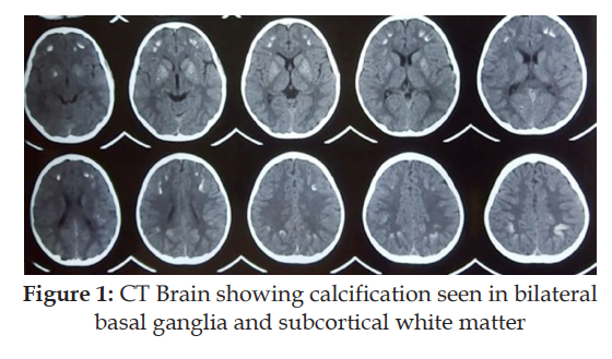
Background: Fahr’s disease is a rare neurological disorder characterized by abnormal calcified deposits in the basal ganglia. Only a small number of pediatric onset cases have been reported in the literature to date, and the majority of the Fahr’s disease literature is based on case reports. Case report: We are reporting a rare case of an 8-year-old child with Fahr’s disease presenting with myoclonus and dysarthria. Bilateral calcifications in the subcortical white matter and basal ganglia were visible on his CT brain. Clinically, he is treated symptomatically and is responding well to clonazepam. Conclusion: We want to highlight that Fahr’s disease can have a wider spectrum of age of onset and also to point out that it should be considered a differential in children presenting with movement disorder with intracerebral calcifications. Our aim is to expand the current knowledge available on clinic-radiological features of this disease entity and encourage further research in genetic and treatment aspects by increasing awareness regarding Fahr’s disease.
References
- 1. Donzuso, G., Mostile, G.,Nicoletti, A. et al. Basal gangliacalcifications (Fahr’s syndrome):related conditions andclinical features. Neurol Sci40, 2251–2263 (2019).
- 2. Fahrs T. Idiopathische Verkalkungender HirngefaBe.Zentrabiatt Allgemeine furPathologie. 1930; 50; 129-133.
- 3. Shafaq S., Hafiz M.A., Maheen A. et al: Fahr’s syndrome: Literature review of current evidence. Orphanet J Rare Dis, 2013; 8: 156
- 4. Akaninyene Asuquo O,Anikwe Jude C., DerekCocker. Fahr’s disease: a rareneurological presentation in atropical setting. Clin CaseRep. 2015 Oct; 3(10): 806–808.
- 5. Oh S.K., Lee J.W., Yoo B.D. et al: Epileptic seizure revealing a Fahr’s syndrome with pseudohypoparathyroidism: A case report. J Korean Soc Emerg Med, 2014; 25(5): 636–40
- 6. Billard C., Dulac O., Boulouche J., Echenne B, et al. (1989) Encephalopathy with Calcifications of the basalganglia in children: areappraisal of Fahr’s syndrome with respect to 14 new cases. Neuropediatrics 20(1): 12-9.
- 7. Gopen Kumar Kundu, et al. A Child with Fahr’s Disease. BAOJ Pediat 2017 3: 2
- 8. Muoneke V Uzoamaka et al. Idiopathic basal ganglia calcification (Fahr’s disease) in a 9-year-old Nigerian child. Niger J Paediatr 2021; 48 (1): 46 – 49
- 9. Sandhu J.K., Kaul V., Sandhu K.K. Fahr’s syndrome presenting as focal seizures in an adolescent: a case report. Int J Contemp Pediatr 2024; 11: 1464-6.
- 10. Sinha R.1, Sodhi K.2, John BM.3, Singh D. Fahr’s Disease: A Case Report. Journal of Nepal Paediatric Society. January-June, 2010/ Vol 30/Issue 1
- 11. Ahad M.A., Bala C., Karim S.: Fahr’s syndrome. Bangladesh Medical Journal Khulna 2013, 45(1–2): 33–35.
- 12. Shafaq Saleem, Hafiz MuhammadAslam. Fahr’s syndrome: literaturereview of current evidence. Orphanet J Rare Dis. 2013; 8: 156.
- 13. Manyam B.V.: What is and what is not ‘Fahr’s disease’. Parkinsonism RelatDisord 2005, 11(2):73–80.
- 14. Rastogi R., Singh A.K., Rastogi U.C., Chander Mohan C., Vaibhav Rastogi V. (2011) Fahr’s syndrome: a rare clinico-radiologic entity. Med J Armed Forces India 67(2): 159-161.
- 15. Sahin N., Solak A., Genc B., Kulu U.: Fahr disease: Use of susceptibility- weighted imaging for diagnostic dilemma with magnetic resonance imaging. Quant Imaging Med Surg, 2015; 5(4): 628–32
Data Sharing Statement
There are no additional data available. All raw data and code are available upon request.
Funding
This research received no funding.
Author Contributions
All authors contributed significantly to the work and approve its publication.
Ethics Declaration
No Ethical issues involved
Acknowledgements
We would like to express our gratitude to the patients, their families, and all those who have contributed to this study.
Conflicts of Interest
No conflicts of interest.
About this article
Cite this article
Prajakta S. Ghatage, Abhijeet S. Ghatage, Sharad T. Ghatage et. al, A Rare Case of Pediatric Fahr’s Disease with Myoclonus as Initial Presentation. International Journal of Neurology and Neurosurgery. 2025; 17(2): 184-187.
Licence:
Attribution-Non-commercial 4.0 International (CC BY-NC 4.0)This license enables reusers to distribute, remix, adapt, and build upon the material in any medium or format for noncommercial purposes only, and only so long as attribution is given to the creator.
| Received | Accepted | Published |
|---|---|---|
| February 21, 2025 | April 25, 2025 | July 30, 2025 |
DOI: 10.21088/ijnns.0975.0223.17225.14
Keywords
Fahr’s diseaseMyoclonusBasal ganglia calcificationSearch for Similar Articles
Similar Articles
- Intramedullary Capillary Hemangioma of Conus Medullaris: Case Report and Review...
- Atypical Miller Fisher Syndrome
- Therapeutic Potential of Passiflora incarnata in the Management of Neurological...
- Central or Axial Atlantoaxial Dislocation: Redefining Instability at the Craniov...
- Assessment of Neurocognitive Functions in Post-Acute Stroke Patients Undergoing...
Article Level Metrics
Last UpdatedThursday 30 July 2026, 10:12:23 (IST)
1464
Accesses
17
244
00
Citations
NA
NA
NA
Download citation
Article Keywords
Keyword Highlighting
Highlight selected keywords in the article text.
Timeline
| Received | February 21, 2025 |
| Accepted | April 25, 2025 |
| Published | July 30, 2025 |
licence
Attribution-Non-commercial 4.0 International (CC BY-NC 4.0)
This license enables reusers to distribute, remix, adapt, and build upon the material in any medium or format for noncommercial purposes only, and only so long as attribution is given to the creator.