Heading
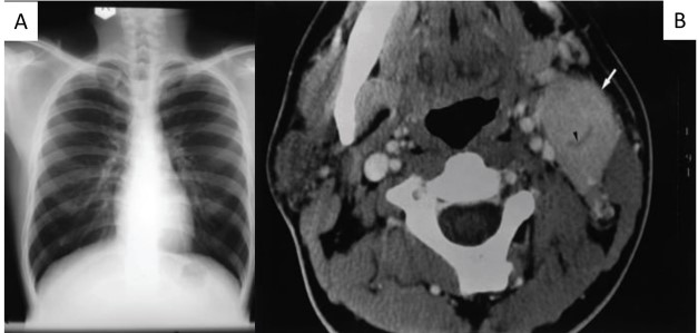
Description: (A) Chest radiograph (posteroanterior view) showing normal lung fields and mediastinal contours, with no
evidence of mediastinal widening, pulmonary lesion, or lymphadenopathy; (B) Axial contrast-enhanced computed
tomography (CT) scan of the neck demonstrating an enlarged, well-enhancing left level II cervical lymph node (white
arrow) with a central crescentic low-attenuation band (arrowhead), a radiological feature suggestive of a hypervascular lymphoid lesion.