Full Text (PDF)
International Physiology 10(1):p 21-27, January-April 2022. | DOI: https://doi.org/10.21088/ip.2347.1506.10122.3
Original Article
Skin Substitutes: An Overview
Ravi Kumar Chittoria, Jacob Antony Chakiath
Author Information
Licence:
Attribution-Non-commercial 4.0 International (CC BY-NC 4.0)This license
enables reusers to distribute, remix, adapt, and build upon the material in any
medium or format for noncommercial purposes only, and only so long as
attribution is given to the creator.
International Physiology 10(1):p 21-27, January-April 2022. | DOI: https://doi.org/10.21088/ip.2347.1506.10122.3
How Cite This Article:
Ravi Kumar Chittoria, Jacob Antony Chakiath/ Skin Substitutes: An Overview/International Physiology.2022;10(1):21–27.Timeline
Received : January 13, 2022
Accepted : March 30, 2022
Published : April 30, 2022
Abstract
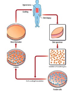
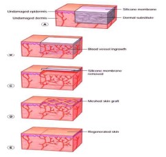
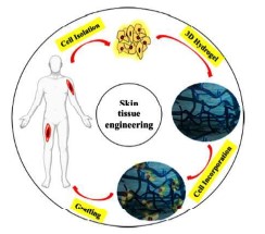
Skin substitutes are a heterogeneous group of biologic, synthetic, or biosynthetic materials that can provide coverage of open skin wounds. The aim of skin substitutes is to replicate the properties of the normal skin. Biocompatibility, antimicrobial activity, appropriate hydrophilicity, and biodegradability are all desirable qualities in a skin substitute. The goal of tissue engineering research is to develop cell-based wound substitutes or wound covers that promote cell migration, differentiation, and vascularization to facilitate wound healing.
References
- 1. Shevchenko, R.V.; James, S.E. A review of tissueengineered skin bioconstructs available for skin reconstruction. J. R. Soc. Interface 2009, 7, 229–258.[CrossRef] [PubMed]
- 2. Rowan, M.P.; Cancio, L.C.; Elster, E.A.; Burmeister, D.M.; Rose, L.F.; Natesan, S.; Chan, R.K.; Christy, R.J.; Chung, K.K. Burn wound healing and treatment: Review and advancements. Crit. Care 2015, 19, 1–12. [CrossRef] [PubMed]
- 3. Klar, A.S.; Michalak-Mi´cka, K.; Biedermann, T.; Simmen-Meuli, C.; Reichmann, E.; Meuli, M. Characterization of M1 and M2polarization of macrophages in vascularized human dermoepidermal skin substitutes in vivo. Pediatr. Surg. Int. 2017, 34, 129–135. [CrossRef]
- 4. Pogorielov, M.; Hapchenko, A.; Pogorielov, O.O.M. Tissue Engineering: Challenges and Selected Application. Adv. Tissue Eng.Regen. Med. Open Access 2017, 3, 1–6.[CrossRef]
- 5. Olena, P.; Prokopyuk, V.; Figueiredo, C.; Pogozhykh, D. Placenta and Placental Derivatives in Regenerative Therapies: Experimental Studies, History, and Prospects. Stem Cells Int. 2018, 2018, 1–14. [CrossRef]
- 6. Maxson, S.; Lopez, E.A.; Yoo, D.; DanilkovitchMiagkova, A.; Leroux, M.A. Concise Review: Role of Mesenchymal Stem Cells in Wound Repair. STEM CELLS Transl. Med. 2012, 1, 142–149. [CrossRef]
- 7. Lavery, L.A.; Fulmer, J.; Shebetka, K.A.; Regulski, M.; Vayser, D.; Fried, D.; Kashefsky, H.; Owings, T.M.; Nadarajah, J. The Grafix Diabetic Foot Ulcer Study Group The efficacy and safety of Grafix® for the treatment of chronic diabetic foot ulcers: Results of a multi-centre, controlled, randomised, blinded, clinical trial. Int. Wound J. 2014, 11, 554–560. [CrossRef] [PubMed]
- 8. Wood, F.M.; Kolybaba, M.; Allen, P. The use of cultured epithelial autograft in the treatment of major burn wounds: Eleven years of clinical experience. Burns 2006, 32, 538–544. [CrossRef] [PubMed]
- 9. Barret, P.J.;Wolf, S.E.; Desai, M.H.; Herndon, D.N. Cost-Efficacy of Cultured Epidermal Autografts in Massive Pediatric Burns. Ann. Surg. 2000, 231, 869–876. [CrossRef]
- 10. Marston, W.A.; Hanft, J.; Norwood, P.; Pollak, R. The Efficacy and Safety of Dermagraft in Improving the Healing of Chronic Diabetic Foot Ulcers: Results of a prospective randomized trial. Diabetes Care 2003, 26, 1701–1705. [CrossRef]
- 11. Hart, C.E.; Loewen-Rodriguez, A.; Lessem, J. Dermagraft: Use in the Treatment of Chronic Wounds. Adv. Wound Care 2012, 1, 138–141.[CrossRef]
- 12. Kumar, S.; Kang, H.J.; Berthiaume, F. Scaffolds for epidermal tissue engineering. In Handbook of Tissue Engineering Scaffolds; Woodhead Publishing: Cambridge, UK, 2019; Volume 2, pp. 173–191.
- 13. Curran, M.P.; Plosker, G.L. Bilayered Bioengineered Skin Substitute (Apligraf®): A Review of Its Use in the Treatment of Venous Leg Ulcers and Diabetic Foot Ulcers. BioDrugs2002, 16, 439–455. [CrossRef] [PubMed]
- 14. Pourmoussa, A.; Gardner, D.J.; Johnson, M.B.; Wong, A.K. An update and review of cell-based wound dressings and their integration into clinical practice. Ann. Transl. Med. 2016, 4, 457. [CrossRef]
- 15. Braziulis, E.; Biedermann, T.; Hartmann-Fritsch, F.; Schiestl, C.; Pontiggia, L.; Böttcher-Haberzeth, S.; Reichmann, E.; Meuli, M.Skingineering I: Engineering porcine dermo-epidermal skin analogues for autologous transplantation in a large animal model.Pediatr. Surg. Int. 2011, 27, 241–247. [CrossRef]
- 16. Schiestl, C.; Neuhaus, K.; Biedermann, T.; BöttcherHaberzeth, S.; Reichmann, E.; Meuli, M. Novel Treatment for Massive Lower Extremity Avulsion Injuries in Children: Slow, but Effective with Good Cosmesis. Eur. J. Pediatr. Surg. 2010, 21, 106–110. [CrossRef]
- 17. Schiestl, C.; Stiefel, D.; Meuli, M. Giant naevus, giant excision, eleg (i) ant closure? Reconstructive surgery with Integra ArtificialSkin® to treat giant congenital melanocytic naevi in children. J. Plast. Reconstr. Aesthet. Surg. 2010, 63, 610–615. [CrossRef]
- 18. Zimoch, J.; Padial, J.S.; Klar, A.S.; Vallmajo-Martin, Q.; Meuli, M.; Biedermann, T.; Wilson, C.J.; Rowan, A.; Reichmann, E. Polyisocyanopeptide hydrogels: A novel thermo-responsive hydrogel supporting pre-vascularization and the development of organotypic structures. ActaBiomater. 2018, 70, 129–139. [CrossRef]
- 19. Tavakoli, S.; Kharaziha, M.; Nemati, S.; Kalateh, A. Nanocomposite hydrogel based on carrageenancoated starch/cellulose nanofibers as a hemorrhage control material. Carbohydr. Polym. 2021, 251, 117013. [CrossRef]
- 20. Tavakoli, S.; Kharaziha, M.; Kermanpur, A.; Mokhtari, H. Sprayable and injectable visible-light Kappa-carrageenan hydrogel for in-situ soft tissue engineering. Int. J. Biol. Macromol. 2019, 138, 590–601. [CrossRef] [PubMed]
- 21. Rana, D.; Kumar, T.S.; Ramalingam, M. Cell-laden hydrogels for tissue engineering. J. Biomater. Tissue Eng. 2014, 4, 507–535.[CrossRef]
- 22. Q.; Mai, Y.-W. Biomaterials for Implants and Scaffolds. In Biomaterials Science and Engineering; Springer: Berlin, Germany, 2017; Volume 8, ISBN 978-3-662-53572-1.
- 23. Klawitter, J.J.; Hulbert, S.F. Application of porous ceramics for the attachment of load bearing internal orthopedic applications. J.Biomed. Mater. Res. 1971, 5, 161–229. [CrossRef]
- 24. Yeh, J.; Blumling, J.; Karp, J.M.; Gantz, J.; Chandawarkar, A.; Eng, G.; Iii, J.B.; Langer, R.; Khademhosseini, A. Micromolding of shapecontrolled, harvestable cell-laden hydrogels. Biomaterials 2006, 27, 5391–5398. [CrossRef] [PubMed]
- 25. Eke, G.; Mangir, N.; Hasirci, N.; MacNeil, S.; Hasirci, V. Development of a UV crosslinked biodegradable hydrogel containing adipose derived stem cells to promote vascularization for skin wounds and tissue engineering. Biomaterials 2017, 129, 188–198. [CrossRef]
- 26. Murphy, S.V.; Atalaa, A. 3D bioprinting of tissues and organs. Nat. Biotechnol. 2014, 32, 773–785. [CrossRef]
- 27. Li, H.; Tan, C.; Li, L. Review of 3D printable hydrogels and constructs. Mater. Des. 2018, 159, 20–38. [CrossRef]
- 28. Hng, G.; Li, F.; Zhao, X.; Ma, Y.; Li, Y.; Min, L.; Jin, G.; Lu, T.J.; Genin, G.M.; Xu, F. Functional and Biomimetic Materials for Engineering of the ThreeDimensional Cell Microenvironment. Chem. Rev. 2017, 117, 12764–12850. [CrossRef] [PubMed]
- 29. Yanez, M.; Rincon, J.; Dones, A.; De Maria, C.; Gonzales, R.; Boland, T. In Vivo Assessment of Printed Microvasculature in a Bilayer Skin Graft to Treat Full-Thickness Wounds. Tissue Eng. Part A2015, 21, 224–233. [CrossRef]
- 30. Albanna, M.; Binder, K.W.; Murphy, S.V.; Kim, J.; Qasem, S.A.; Zhao, W.; Tan, J.; El-Amin, I.B.; Dice, D.D.; Marco, J.; et al. In SituBioprinting of Autologous Skin Cells Accelerates Wound Healing of Extensive Excisional Full-Thickness Wounds. Sci. Rep. 2019,9, 1–15. [CrossRef] [PubMed]
- 31. Hafezi, F.; Scoutaris, N.; Douroumis, D.; Boateng, J.S. 3D printed chitosan dressing crosslinked with genipin for potential healingof chronic wounds. Int.J. Pharm. 2019, 560, 406–415. [CrossRef]
- 32. Admane, P.; Gupta, A.C.; Jois, P.; Roy, S.; Lakshmanan, C.C.; Kalsi, G.; Bandyopadhyay, B.; Ghosh, S. Direct 3D bioprintedfullthicknessskin constructs recapitulate regulatory signaling pathways and physiology of human skin. Bioprinting2019, 15, e00051.[CrossRef]
Data Sharing Statement
There are no additional data available. All raw data and code are available upon request.
Funding
This research received no funding.
Author Contributions
All authors contributed significantly to the work and approve its publication.
Ethics Declaration
This article does not involve any human or animal subjects, and therefore does not require ethics approval.
Acknowledgements
We would like to express our gratitude to the patients, their families, and all those who have contributed to this study.
Conflicts of Interest
No conflicts of interest in this work.
About this article

Cite this article
Ravi Kumar Chittoria, Jacob Antony Chakiath/ Skin Substitutes: An Overview/International Physiology.2022;10(1):21–27.
Licence:
Attribution-Non-commercial 4.0 International (CC BY-NC 4.0)This license
enables reusers to distribute, remix, adapt, and build upon the material in any
medium or format for noncommercial purposes only, and only so long as
attribution is given to the creator.
| Received | Accepted | Published |
|---|---|---|
| January 13, 2022 | March 30, 2022 | April 30, 2022 |
DOI: https://doi.org/10.21088/ip.2347.1506.10122.3
Keywords
Skin substitute.Search for Similar Articles
Similar Articles
- Mechanisms of Myocardial Damage and Structural Remodeling Caused by Doxorubicin-...
- Impact of Sleep Deprivation on Human Physiology: A Review
- The Effect of a Brief Heart Rate Variability Biofeedback Session on Salivary Cor...
- Impact of Vitamin D Supplementation in Enhancing Pulmonary Function in Adults wi...
- Mona Bedi - Editiorial
Article Level Metrics
Last UpdatedSunday 27 September 2026, 07:35:06 (IST)
1960
Accesses
11
194
00
Citations
NA
NA
NA
Download citation
Article Keywords
Keyword Highlighting
Highlight selected keywords in the article text.
Timeline
| Received | January 13, 2022 |
| Accepted | March 30, 2022 |
| Published | April 30, 2022 |
licence
Attribution-Non-commercial 4.0 International (CC BY-NC 4.0)
This license
enables reusers to distribute, remix, adapt, and build upon the material in any
medium or format for noncommercial purposes only, and only so long as
attribution is given to the creator.