Full Text (PDF)
Indian Journal of Preventive Medicine 13(2):p 69-73, July-Dec 2025. | DOI: 10.21088/ijpm.2321.5917.13225.4
Case Report
Rhinosporidiosis in A 10-Year-Old Child: A Case Report Highlighting the Environmental and Public Health Implications of Aquatic Spore Exposure
Rajeev Aravindakshan, Vivekanand Ashok, Deepa Appu, Smriti Suresh
Author Information
Licence:
Attribution-Non-commercial 4.0 International (CC BY-NC 4.0)This license enables reusers to distribute, remix, adapt, and build upon the material in any medium or format for noncommercial purposes only, and only so long as attribution is given to the creator.
Indian Journal of Preventive Medicine 13(2):p 69-73, July-Dec 2025. | DOI: 10.21088/ijpm.2321.5917.13225.4
How Cite This Article:
Vivekanand A, Appu D, Suresh S, et al. Rhinosporidiosis in A 10-Year-Old Child: A Case Report Highlighting The Environmental and Public Health Implications of Aquatic Spore Exposure. J Prev Med. 2025;13(2):69-73.Timeline
Received : July 22, 2025
Accepted : September 27, 2025
Published : December 20, 2025
Abstract
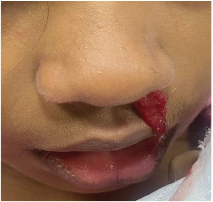
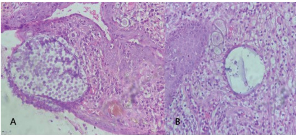
Rhinosporidiosis, a chronic granulomatous infection that can be prevented and is mainly caused by community practices such as bathing in contaminated ponds, is endemic in places like Tamil Nadu, India. The treatment of a 10-year-old girl from Palakkad, Kerala, who presented with a reddish mass jutting from her left nasal cavity resulting in nasal obstruction and epistaxis, is detailed in this case report. Contrast-enhanced CT scan revealed a nasal mass with reduced central density was discovered at the inferior turbinate. An endoscopic nasal polypectomy with cauterization was performed. Rhinosporidiosis with distinctive sporangia was confirmed by histopathological examination. The higher prevalence of Rhinosporidium seeberi in neighbouring Tamil Nadu raises the possibility of exposure through pond bathing, even though Palakkad’s ponds are not officially recognized as reservoirs for the disease. This case emphasizes the preventable nature of rhinosporidiosis and the effectiveness of combined surgical and medical management through public health initiatives in endemic areas.
References
- 1. Töz S. Rhinosporidium seeberi: Is It a Fungi or Parasite?. Turkiye Parazitol Derg. 2020;44(4):258-260.
- 2. Rajendran S, Ashok SS, Ramkumar T. Primary nasal rhinosporidiosis - a ten year multicentre experience: what we know?. Int J Otorhinolaryngol Head Neck Surg 2020;6:343-6.
- 3. Chandran M, Mehta R, Nagarkar NM, Bhargava A, Mohapatra E, Pati SK. R h i n o s p o r i d i o s i s - E p i d e m i o l o g i c a l , Clinicoradiological, Immunological Profile. Iran J Otorhinolaryngol. 2023 Sep;35(130): 255-262.
- 4. Mathew S, Arora RD, Prabha N, Kamble P, Satpute SS, Nagarkar NM. Retroanalytical Study of Epidemiological Factors of Rhinosporidiosis. Int Arch Otorhinolaryngol. 2020 Nov 30;25(4):e504-e508.
- 5. Salim T, Komu F. Varied Presentations of Cutaneous Rhinosporidiosis: A Report of Three Cases. Indian J Dermatol. 2016 MarApr;61(2):209-12.
- 6. Madana J, Yolmo D, Gopalakrishnan S, Saxena SK. Laryngotracheal rhinosporidiosis. Ear Nose Throat J. 2013 Jul;92(7):E27-30.
- 7. Alam MS, Shrirao N. Clinical Spectrum and Management Outcome of Ocular and Adnexal Rhinosporidiosis. J Curr Ophthalmol. 2022 Nov 30;34(3):341-346.
- 8. Krishnamoorthi, Sivanantham. Rhinosporidiosis. Tropical Parasitology. Jan–Jun 2025;15(1): p 54-55.
- 9. Arias AF, Romero SD, Garcés CG. Case Report: Rhinosporidiosis Literature Review. Am J Trop Med Hyg. 2020 Dec 7;104(2):708-711.
- 10. Raja K, Thangavel S, Kushwaha A, Srinivas BH, Kar R, Alexander A. Management of Disseminated Rhinosporidiosis: Experience From a Single Tertiary Institution. Turk Arch Otorhinolaryngol. 2024 Oct 23;62(2):66-71. doi: 10.4274/tao.2023.2022-9-5.
- 11. Doddawad VG, Singh R, S S. A new technique to resolve Nasal Rhinosporidiosis: A case report with review of literature. Int J Surg Case Rep. 2022;92:106807.
- 12. Shariff, M. A. (2018). A clinicopathological study of rhinosporidiosis in a tertiary care hospital. International Journal of Otorhinolaryngology and Head and Neck Surgery, 4(4), 981–985.
Data Sharing Statement
There are no additional data available.
Funding
This research received no funding.
Author Contributions
All authors contributed significantly to the work and approve its publication.
Ethics Declaration
This article does not involve any human or animal subjects, and therefore does not require ethics approval.
Acknowledgements
Information not provide.
Conflicts of Interest
The authors report no conflicts of interest in this work.
About this article
Cite this article
Vivekanand A, Appu D, Suresh S, et al. Rhinosporidiosis in A 10-Year-Old Child: A Case Report Highlighting The Environmental and Public Health Implications of Aquatic Spore Exposure. J Prev Med. 2025;13(2):69-73.
Licence:
Attribution-Non-commercial 4.0 International (CC BY-NC 4.0)This license enables reusers to distribute, remix, adapt, and build upon the material in any medium or format for noncommercial purposes only, and only so long as attribution is given to the creator.
| Received | Accepted | Published |
|---|---|---|
| July 22, 2025 | September 27, 2025 | December 20, 2025 |
DOI: 10.21088/ijpm.2321.5917.13225.4
Keywords
RhinosporidiosisAquatic ExposureSporangiaPolypectomyDapsoneSearch for Similar Articles
Similar Articles
- Recent Advances in Diabetes and Obesity Management
- Forensic Implications of Clinically Significant Diseases: From Sudden Cardiac De...
- Mindful Eating for Restorative Sleep: A Community-Based Approach to Sleep Hygien...
- Utilizing AI for Identifying and Addressing Health Disparities in Communities: A...
- The Importance of Super Oxidised Solution in the Management of Thermal Burn Wou...
Article Level Metrics
Last UpdatedTuesday 07 July 2026, 05:13:42 (IST)
604
Accesses
18
146
00
Citations
NA
NA
NA
Download citation
Article Keywords
Keyword Highlighting
Highlight selected keywords in the article text.
Timeline
| Received | July 22, 2025 |
| Accepted | September 27, 2025 |
| Published | December 20, 2025 |
licence
Attribution-Non-commercial 4.0 International (CC BY-NC 4.0)
This license enables reusers to distribute, remix, adapt, and build upon the material in any medium or format for noncommercial purposes only, and only so long as attribution is given to the creator.