Full Text (PDF)
International Journal of Neurology and Neurosurgery 15(2):p 47-53, April – June 2023. | DOI: 10.21088/ijnns.0975.0223.15223.2
Original Article
Retrospective Observational Study of Clinical Profile, Neurosurgical Management and Outcome of Intracranial Tumors in a Tertiary Care Government Teaching Hospital
Author Information
Licence:
Attribution-Non-commercial 4.0 International (CC BY-NC 4.0)This license enables reusers to distribute, remix, adapt, and build upon the material in any medium or format for noncommercial purposes only, and only so long as attribution is given to the creator.
International Journal of Neurology and Neurosurgery 15(2):p 47-53, April – June 2023. | DOI: 10.21088/ijnns.0975.0223.15223.2
How Cite This Article:
Mishra A. Retrospective observational study of clinical profile, neurosurgical management and outcome of intracranial tumors in a tertiary care government teaching hospital. Int J Neurol Neurosurg. 2023;15(2):47–53.Timeline
Received : March 30, 2023
Accepted : April 28, 2023
Published : May 30, 2023
Abstract
Introduction: A brain tumor, known as an intracranial tumor, is an abnormal mass of tissue in which cells grow and multiply uncontrollably, seemingly unchecked by the mechanisms that control normal cells. The two main groups of brain tumors are termed primary and metastatic.
Aim: The aim is to study the different causes, clinical outcome and management of the patient.
Materials and Methods: This study is a retrospective observational study and included all the patient who underwent surgery for intracranial tumor in our hospital from December 2016 to December 2021.
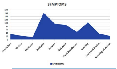
Results: Total 350 patients underwent the surgery for intracranial tumor who was admitted in our hospital from December 2016 to December 2021. In all the patient presented headache and seizures was the commonest symptom.
Conclusion: In our retrospective study conducted in 350 patients, headache was the most common symptom and more frequently associated with males and cerebral convexity is the most common location of occurrence with meningioma being the most common.
References
- 1. Counsell CE, Grant R. Incidence studies of primary and secondary intracranial tumors: a systematic review of their methodology and results. J Neurooncol. 1998;37(3):241-50.
- 2. Niederhuber JE, Armitage JO, Kastan MB, Doroshow JH, Tepper JE, editors. Cancer of the central nervous system. In: Abeloff’s clinical oncology. 6th ed. Philadelphia: Elsevier; 2020.
- 3. Ostrom QT, Gittleman H, Farah P, Ondracek A, Chen Y, Wolinsky Y, et al. CBTRUS statistical report: primary brain and central nervous system tumours diagnosed in the United States in 2006-2010. Neuro Oncol. 2013;15 Suppl 2:ii1-56.
- 4. Burger PC, Scheithauer BW. Tumours of the central nervous system. In: AFIP atlas of tumour pathology. Washington (DC): Armed Forces Institute of Pathology; 2007.
- 5. Adesina AM, Tihan T, Fuller CE, Poussaint TY, editors. Atlas of pediatric brain tumors. New York: Springer; 2009.
- 6. Baker GJ, Yadav VN, Motsch S, Koschmann C, Calinescu AA, Mineharu Y, et al. Mechanisms of glioma formation: iterative perivascular glioma growth and invasion leads to tumor progression, VEGF independent vascularization, and resistant and antiangiogenic therapy. Neoplasia. 2014;16(7):543-61.
- 7. Singh SK, Hawkins C, Clarke ID, Squire JA, Bayani J, Hide T, et al. Identification of human brain tumour initiating cells. Nature. 2004;432(7015):396-401.
- 8. Louis DN, Perry A, Reifenberger G, von Deimling A, Figarella-Branger D, Cavenee WK, et al. The 2016 World Health Organization classification of tumors of the central nervous system: a summary. Acta Neuropathol. 2016;131(6):803-20.
- 9. Ohgaki H, Wiestler OD, Cavenee WK, editors. WHO classification of tumours of the central nervous system. 4th ed. Lyon (France): International Agency for Research on Cancer; 2007.
- 10. Van Meir EG, editor. CNS cancer: models, markers, prognostic factors, targets, and therapeutic approaches. New York: Springer; 2009.
- 11. Thomas B, Krishnamoorthy T, Radhakrishnan VV, Kesavadas C. Moving closer to pathology and pathophysiology. Neuroradiology. 2007;49(9):733-8.
- 12. Vivas AC, Reintjes S, Shimony N, Vale FL. Surgery for amygdala and uncus: a case series of glioneuronal tumors. Acta Neurochir (Wien). 2020;162(4):795-801.
- 13. Gregg N, Arber A, Ashkan K, Brazil L, Bhangoo R, Beaney R, et al. Neurobehavioural changes in patients following brain tumour: patients and relatives perspective. Support Care Cancer. 2014;22(11):2965–72.
- 14. Park BJ, Kim HK, Sade B, Lee JH. Epidemiology. In: Lee JH, editor. Meningiomas: diagnosis, treatment, and outcome. London: Springer; 2009.
- 15. Maier SE, Sun Y, Mulkern RV. Diffusion imaging of brain tumors. NMR Biomed. 2010;23(7):849–64.
- 16. Cha S. Perfusion MR imaging of brain tumors. Top Magn Reson Imaging. 2004;15(5):279–89.
- 17. Bullitt E, Jung I, Muller K, Gerig G, Aylward S, Joshi S, et al. Determining malignancy of brain tumors by analysis of vessel shape. In: Barillot C, Haynor DR, Hellier P, editors. Medical Image Computing and Computer-Assisted Intervention – MICCAI 2004. Berlin, Heidelberg: Springer; 2004. p. 645–53.
- 18. Chamberlain MC, Kormanik PA. Practical guidelines for the treatment of malignant gliomas. West J Med. 1998;168(2):114
Data Sharing Statement
There are no additional data available. All raw data and code are available upon request.
Funding
This research received no funding.
Author Contributions
Whether all authors contributed significantly to the work and approve its publication.
Ethics Declaration
This article does not involve any human or animal subjects, and therefore does not require ethics approval.
Acknowledgements
We would like to express our gratitude to the patients, their families, and all those who have contributed to this study.
Conflicts of Interest
The authors report no conflicts of interest in this work.
About this article

Cite this article
Mishra A. Retrospective observational study of clinical profile, neurosurgical management and outcome of intracranial tumors in a tertiary care government teaching hospital. Int J Neurol Neurosurg. 2023;15(2):47–53.
Licence:
Attribution-Non-commercial 4.0 International (CC BY-NC 4.0)This license enables reusers to distribute, remix, adapt, and build upon the material in any medium or format for noncommercial purposes only, and only so long as attribution is given to the creator.
| Received | Accepted | Published |
|---|---|---|
| March 30, 2023 | April 28, 2023 | May 30, 2023 |
DOI: 10.21088/ijnns.0975.0223.15223.2
Keywords
Intracranial TumorsPrognosisHeadacheMeningiomaGlial TumoursSearch for Similar Articles
Similar Articles
- Intramedullary Capillary Hemangioma of Conus Medullaris: Case Report and Review...
- Atypical Miller Fisher Syndrome
- Therapeutic Potential of Passiflora incarnata in the Management of Neurological...
- Central or Axial Atlantoaxial Dislocation: Redefining Instability at the Craniov...
- Assessment of Neurocognitive Functions in Post-Acute Stroke Patients Undergoing...
Article Level Metrics
Last UpdatedTuesday 29 September 2026, 15:22:30 (IST)
2213
Accesses
32
275
00
Citations
NA
NA
NA
Download citation
Article Keywords
Keyword Highlighting
Highlight selected keywords in the article text.
Timeline
| Received | March 30, 2023 |
| Accepted | April 28, 2023 |
| Published | May 30, 2023 |
licence
Attribution-Non-commercial 4.0 International (CC BY-NC 4.0)
This license enables reusers to distribute, remix, adapt, and build upon the material in any medium or format for noncommercial purposes only, and only so long as attribution is given to the creator.