Full Text (PDF)
Journal of ENT and Allied Sciences 10(1):p 25-28, Jan-June 2025. | DOI: https://doi.org/10.21088/rjeas.2456.5024.10125.4
Case Report
Huge Frontal Sinus Mucocele in an Immunocompromised Patient
Garima Sinha, Avinash Kumar, Mansi Sharma, Ekta Yadav, L. Rimasri Devi
Author Information
Licence:
Attribution-Non-commercial 4.0 International (CC BY-NC 4.0)This license enables reusers to distribute, remix, adapt, and build upon the material in any medium or format for noncommercial purposes only, and only so long as attribution is given to the creator.
Journal of ENT and Allied Sciences 10(1):p 25-28, Jan-June 2025. | DOI: https://doi.org/10.21088/rjeas.2456.5024.10125.4
How Cite This Article:
Avinash Kumar, Garima Sinha, Mansi Sharma et. al, Huge Frontal Sinus Mucocele in an Immunocompromised Patient. RFP J ENT Allied Sci 2025; 10(1): 25–28.Timeline
Received : March 27, 2025
Accepted : April 23, 2025
Published : June 25, 2025
Abstract
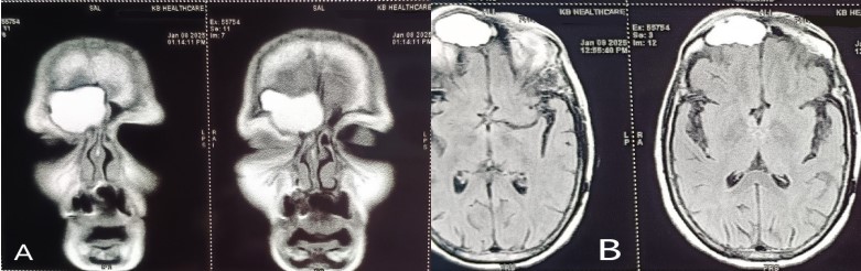
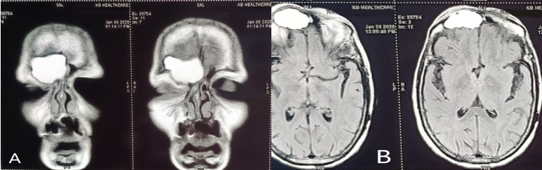
Mucoceles of the paranasal sinuses are defined as cystic expansile lesions (Lageback in 1820). Rollet in 1909 coined the term mucocele. Mucocelesare most commonly found in the frontal sinus (60–65%) butcan also occur in ethmoidsinuses (around 25%), sphenoid sinus (1–2%) and maxillary sinus (10%). The etiopathogenesis of PNS mucocele is multifactorial with most common causes being allergy, inflammation and trauma or less commonly it can be secondary to neoplastic lesions in PNS which are obstructing the sinus ostia.
References
- 1. Alberti PW, Marshall HF, Munro Black JI. Fronto-ethmoidal Mucocele as a cause of Unilateral Proptosis. Br J Ophthalmol. 1968;52:833. doi: 10.1136/bjo.52.11.833.
- 2. Tan CS, Yong VK, Yip LW, Amritj S. An unusual presentation of a giant frontal sinus mucocele manifesting with a subcutaneous forehead mass. Ann Acad Med Singapore. 2005;34:397–8.
- 3. Weitzel EK, Hollier LH, Calzada G, Manolidis S. Single stage management of complex fronto-orbital mucoceles. J Craniofac Surg. 2002;13:739–45. doi: 10.1097/00001665- 200211000-00004.
- 4. Corey W, Chandra RK, Cohen N. Orbital mucopyocele after the use of alloplastic materials in the management of frontal sinus fractures. Otolaryngology. 2006;135:974–976. doi: 10.1016/j.otohns.2005.09.011.
- 5. Edelman RR, Hesselink JR, Zlatkin MB, Crues JV. Clinical Magnetic Resonance Imaging. 3rd ed. Philadelphia: Elsevier; 2006. pp. 2035–7.
- 6. Chobillion MA, Jankowski R. Relationship between mucoceles, nasal polyposis and nasalisation. Rhinology. 2004;43:219–24.
- 7. Rinehart GC, Jackson IT, Potparic Z, Tan RG, Chambers PA. Management of locally aggressive sinus disease using craniofacial exposure and the galealfrontalis fascia-muscle flap. Plast Reconstr Surg 1993;92:1219-26.
- 8. Molteni G, Spinelli R, Panigatti S, Colombo L, Ronchi P. Voluminous frontoethmoidal mucocele with epidural involvement. Surgical treatment by coronal approach. Acta OtorhinolaryngolItal 2003;23:185-90.
- 9. Suri A, Mahapatra AK, Gaikwad S, Sarkar C. Giant mucoceles of the frontal sinus: a series and review. J ClinNeurosci 2004;11:214-8. 7. Avery G, Tang RA, Close LG. Ophthalmic manifestations of mucoceles. Ann Ophthalmol 1983;15:734-7.
- 10. Har-el G. Telescopic extracranial approach to frontal mucoceles with intracranial extension. J Otolaryngol 1995;24:98-101.
- 11. Chew YK, Noorizan Y, Khir A, BritoMutunayagam S, Prepageran N. Frontal mucocoele secondary to nasal polyposis: an unusual complication. Singapore Med J. 2009;50:374–5.
- 12. Lund VJ. Endoscopic management of paranasal sinus mucoceles. J Laryngol Otol. 1998;112:36– 40. doi: 10.1017/s0022215100139854.
Data Sharing Statement
There are no additional data available. All raw data and code are available upon request.
Funding
This research received no funding.
Author Contributions
All authors contributed significantly to the work and approve its publication.
Ethics Declaration
This article does not involve any human or animal subjects, and therefore does not require ethics approval.
Acknowledgements
We would like to express our gratitude to the patients, their families, and all those who have contributed to this study.
Conflicts of Interest
No conflicts of interest in this work.
About this article

Cite this article
Avinash Kumar, Garima Sinha, Mansi Sharma et. al, Huge Frontal Sinus Mucocele in an Immunocompromised Patient. RFP J ENT Allied Sci 2025; 10(1): 25–28.
Licence:
Attribution-Non-commercial 4.0 International (CC BY-NC 4.0)This license enables reusers to distribute, remix, adapt, and build upon the material in any medium or format for noncommercial purposes only, and only so long as attribution is given to the creator.
| Received | Accepted | Published |
|---|---|---|
| March 27, 2025 | April 23, 2025 | June 25, 2025 |
DOI: https://doi.org/10.21088/rjeas.2456.5024.10125.4
Keywords
MucoceleFrontal SinusFessDrafSearch for Similar Articles
Similar Articles
- Hidden Dangers of Oral Hygiene: Oropharyngeal Impalement by Toothbrush in a 3-Ye...
- Pseudomonas Aeruginosa in an Immunocompetent Individual with Acute Tonsillitis;...
- Functional Outcome of Abbe–Estlander Flap in Lower Lip Carcinoma with Commissura...
- Primary Laryngeal Histoplasmosis
- Orignal Efficacy and Safety of Balloon Catheter Dilation for Pediatric Chronic R...
Article Level Metrics
Last UpdatedSunday 27 September 2026, 06:10:33 (IST)
1050
Accesses
4
157
00
Citations
NA
NA
NA
Download citation
Article Keywords
Keyword Highlighting
Highlight selected keywords in the article text.
Timeline
| Received | March 27, 2025 |
| Accepted | April 23, 2025 |
| Published | June 25, 2025 |
licence
Attribution-Non-commercial 4.0 International (CC BY-NC 4.0)
This license enables reusers to distribute, remix, adapt, and build upon the material in any medium or format for noncommercial purposes only, and only so long as attribution is given to the creator.