Heading
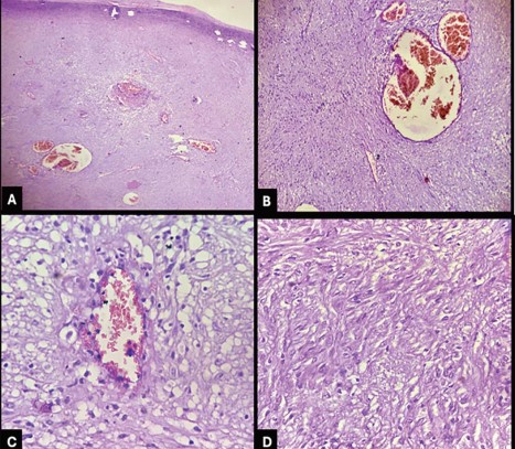
Description: Microphotographs of the tumor show cellular lesion comprising of smooth muscle cells showing interlacing patterns with collagen fibres in between and presence of several thin walled dilated congested vascular channels between the fascicles. (H and E stain a. 10X; b. 10X; c. 40x; d. 40X)