Heading
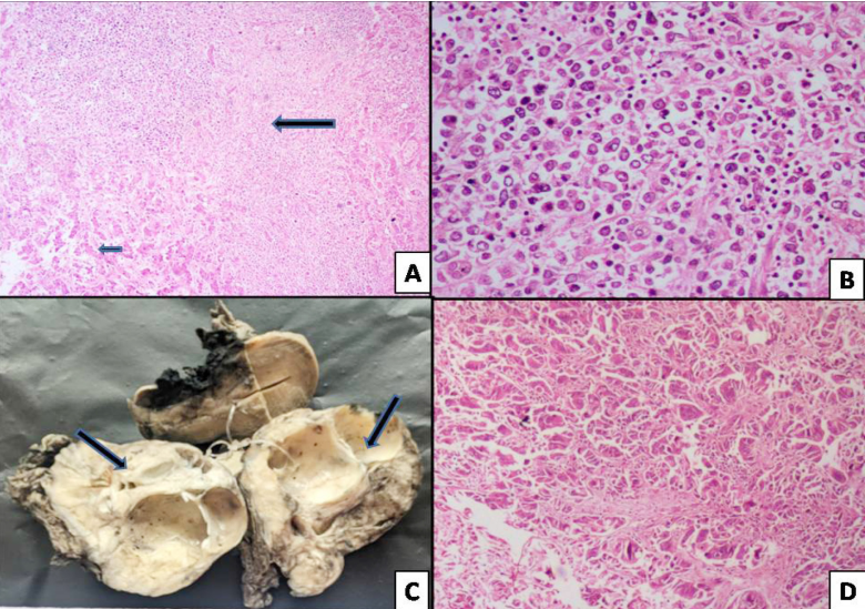
Description: Fig. 2: A) Non-Hodgkin’s lymphoma (long arrow) involving the liver parenchyma (short arrow). B) Diffuse infiltration of large Non-Hodgkin’s lymphoma cells. C) Gross image showing metastatic ovarian malignancy (arrows). D) Cohesive and dyscohesive metastatic tumor cells involving the ovarian parenchyma. (H&E stain; A- x5; B- x20; D- x10).