Full Text (PDF)
Indian Journal of Forensic Odontology 17 (2):p 58-66, July - Dec 2024. | DOI: https://doi.org/10.21088/ijfo.0974.505X.17124.3
Original Article
Gender Determination by Morphometric Analysis of Maxillary Sinus, Orbital Index and Foramen Magnum: A Retrospective CBCT Study
Padmanthan Renganathan, Harsha Vardhan B.G., Saraswathi K Gopal
Author Information
Licence:
Attribution-Non-commercial 4.0 International (CC BY-NC 4.0)This license enables reusers to distribute, remix, adapt, and build upon the material in any medium or format for noncommercial purposes only, and only so long as attribution is given to the creator.
Indian Journal of Forensic Odontology 17 (2):p 58-66, July - Dec 2024. | DOI: https://doi.org/10.21088/ijfo.0974.505X.17124.3
How Cite This Article:
Padmanthan Renganathan, Harsha Vardhan.B.G., Saraswathi. K. Gopal et al. Gender Determination by Morphometric Analysis of Maxillary Sinus, Orbital Index and Foramen Magnum: A Retrospective CBCT Study. Ind J Forensic Odontol 2024;17(1):58-66.Timeline
Received : July 06, 2024
Accepted : September 07, 2024
Published : December 25, 2024
Abstract
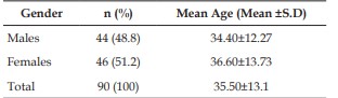
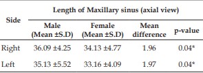
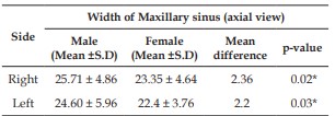
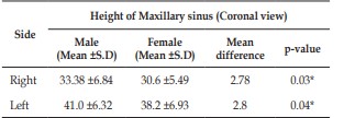
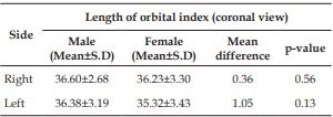
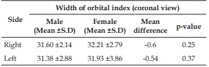
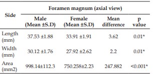
Background: Gender determination is a crucial aspect of forensic anthropology, playing a vital role in the identification process of unknown human remains. Recent advancements in imaging technologies, particularly Cone Beam Computed Tomography (CBCT), have the way for more accurate and non-invasive methods of gender estimation through morphometric analysis.1 The maxillary sinus, orbital index, and foramen magnum are anatomical structures that exhibit sexual dimorphism, meaning their dimensions differ between males and females.1 The maxillary sinus, located within the maxillary bone, shows distinct variations in size and shape based on gender. Similarly, the orbital index, which is the ratio of the orbital height to its width, varies significantly between the gender.2 The foramen magnum, a crucial structure at the base of the skull, also presents measurable differences in size and shape between males and females.3 This retrospective study aims to evaluate the efficacy of using CBCT to determine gender through the morphometric analysis of these anatomical structures. Aim and Objectives: Aim of the study is to determine gender using morphometric analysis of maxillary sinus, orbital index &foramen magnum using CBCT. Objectives of the study is to evaluate the morphometric analysis of maxillary sinus of height, width and depth, morphometric analysis of orbital index of height and width and to evaluate the morphometric analysis of foramen magnum of length width and shape. Methodology: 90 CBCT images of patients were taken from the department database. The DICOM data will be imported to ROMEXIS 6.1.0 software. The age group selected between 13 and above. Maxillary Sinus: Axial view: a) The width was the longest distance perpendicular from the medial wall of the sinus to the most lateral wall of the lateral process of maxillary sinus in axial view. b) The depth was defined as the longest distance from the most anterior point to the most posterior point of the medial wall in the axial view. In coronal view a) The height was measured away from the inner surface of the anterior border of maxillary sinus and was defined as the longest distance from the lowest point of the sinus floor to the highest point of sinus roof in the coronal view. Orbital Index: In coronal view a) Orbital height: Highest distance between the superior and inferior orbital margin. b) Orbital width: Maximum lateral curved distance between the medial margin and lateral margin of the orbit. Foramen Magnum: a) In axial view the anteroposterior dimension and transverse dimension are measured from the most prominent point. b) The shape of the foramen magnum determined based on the Richards and Jabbour classification. c) Shape of the magnum was found to be circle, semicircle, wide oval, heart, bipointed, birounded, venterally rounded and dorsally convergent. Results: There were about 48.8% of males and 51.2% of females in the study. there is a significant difference in length, height and width of maxillary sinus between males and females and hence, maxillary sinus can be used as an effective parameter in gender determination. The length of orbital index was more in males than females and width of orbital index was less in males than females the mean differences between the gender was found to be statistically nonsignificant. The mean length and width of foramen magnum was more in males than females. This mean difference was found to be statistically significant (p-value=0.01), indicating foramen magnum’s length, width and area can be used as an effective parameter in sex determination. Conclusion: CBCT-based morphometric analysis of the maxillary sinus, orbital index, and foramen magnum is a reliable method for gender determination. This approach shows great potential for enhancing forensic anthropological practices and assisting in the identification of human remains. Future research should focus on validating these findings across larger and more diverse populations and exploring additional cranial landmarks to further improve the robustness of gender determination methods.
References
- 1. Auerkari E. Recent trends in dental forensics. Indones J Leg Forensic Sci. 2008;1:5–12. [Google Scholar].
- 2. Singh NN, Gowhar O, Ain TS, Sultan S. Exploring trends in forensic odontology. J Clin Diagn Res. 2014;8:ZC28–30. [PMC free article] [PubMed] [Google Scholar].
- 3. Balachander N, Babu NA, Jimson S, Priyadharsini C, Masthan KM. Evolution of forensic odontology: An overview. J Pharm Bioallied Sci. 2015;7(Suppl 1):S176–80. [PMC free article] [PubMed] [Google Scholar
- 4. Zakirulla M, Meer A. Modern tools in forensic dentistry. J Contemp Dent. 2011;2:28–32. [Google Scholar].
- 5. Jain S, Singh K, Gupta M, Bagri G, Vashistha DK, Soangra R. Role of forensic odontology in human identification: A review Int J Appl Dent Sci. 2020;6:109–11.
- 6. Jeddy N, Ravi S, Radhika T. Current trends in forensic odontology J Forensic Dent Sci. 2017;9:115–9.
- 7. Botwe BO, Sule DS, Ismael AM et al. Radiologic evaluation of orbital Index among Ghanaians using CT scan. DOI: 10.1186/s40101-017-0145-7
- 8. Lal N, Cornwall J, Dias GJ. Orbital indices in a modern Sinhalese Sri Lankan population. Anatomy 2016;10:205-10.
- 9. Uthman AT, Al-Rawi NH, Al-Timimi JF. Evaluation of foramen magnum in gender determination using helical CT scanning. Dentomaxillofac Radiol. 2012; 41(3):197- 202.
- 10. Nagare SP, Chaudhari RS, Birangane RS, Parkarwar PC. Sex determination in forensic identification, a review. J Forensic Dent Sci 2018;10:61-6.
- 11. Tambawala SS, Karjodkar FR, Sansare K, Prakash N, Dora AC. Sexual dimorphism of foramen magnum using cone beam computed tomography. J Forensic Leg Med 2016;44:29-34.
- 12. Lenza Mg, Lenza Mm, Dalstra M, Melsen B, Cattaneo Pm. An analysis of different approaches to the assessment of upper airway morphology: a CBCT study. Orthod Craniofac Res. 2010; 13:96-105.
- 13. Arora et al., Role of forensic odontology in the Indian Armed Forces: An unexplored arena. J Forensic Dent Sci. 2016 Sep-Dec; 8(3): 173.
- 14. Kurland M. Irrefutable Evidence: Adventures in the History of Forensic Science. Rowman & Littlefield; 2009.
- 15. Lewis JA. Forensic document examination: fundamentals and current trends. Elsevier; 2014 Jan 31.
- 16. Balachander N, Babu NA, Jimson S, Priyadharsini C, Masthan KM. Evolution of forensic odontology: An overview. Journal of pharmacy & bioallied sciences. 2015 Apr;7 Suppl 1):S176.
- 17. Rai B, Kaur J. Forensic odontology: History, scope, and limitations. InEvidence-Based Forensic Dentistry 2013 (pp. 1-7). Springer,Berlin, Heidelberg
- 18. Krishan K, Kanchan T, Garg AK. Dental evidence in forensic identification–An overview, methodology and present status. The open dentistry journal. 2015;9:25
- 19. Amedo O. The role of dentist in the identification of the victims of the catastrophe of the ― Bazar de la Charite , Paris, 4th May, 1897. Dent Cosm.
- 20. Highfield R. London: Daily Telegraph; 1999. Dental detective works gets to the root of Hitler mystery.
- 21. Eckert WG, Garland N. The history of the forensic applications in radiology. Am J Forensic Med Pathol 1984; 5:53-62
- 22. Vanita Rathod, Veena Desai, Siddharth Pundir, Sudhanshu Dixit, and Rashmi Chandraker; Role of forensic dentistry for dental practitioners: A comprehensive study; J Forensic Dent Sci. 2017 MayAug; 9(2): 108–109.
- 23. : Dineshkumar T. Role of Forensic Odontologist in Dentistry. Oral Maxillofac Pathol J 2017;8(2): 136-138.
- 24. Lake AW, James H, Berketa JW. Disaster victim identification: quality management from an odontology perspective. Forensic Sci Med Pathol 2012 June;8(2):157-163.
- 25. Datta P, Sood S, Rastogi P, Bhargava K, Bhargava D, Yadav M. DNA profiling in forensic dentistry. J Indian Acad Forensic Med 2012 AprilJune;34(2):156-159.
- 26. Murakami H, Yamamoto Y, Yoshitome K, Ono T, Okamoto O, Shigeta Y, Doi Y, Miyaishi S, Ishizu H.Forensic study of sex determination using PCR on teeth samples. Acta Med Okayama 2000 Feb;54(1):21-32.
- 27. Shkrum MJ, Ramsay DA. Forensic pathology of trauma. Springer Science & Business Media; 2007 Nov 6.
- 28. Devadiga A. What’s the deal with dental records for practicing dentists? Importance in general and forensic dentistry. J Forensic Dent Sci 2014;6:9-15.
- 29. Madi HA, Swaid S, Al-Amad S. Assessment of the uniqueness of human dentition. J Forensic Odontostomatol 2013;31:30-9.
- 30. Pinchi V, Zei G. Two positive identifications assessed with occasional dental findings on non-dental x-rays. J Forensic Odontostomatol 2008;26:34-8.
- 31. Charangowda BK. Dental records: An overview. J Forensic Dent Sci 2010;2:5-10.
- 32. Avon SL. Forensic odontology: The roles and responsibilities of the dentist. J Can Dent Assoc 2004;70:453-8.
- 33. Dawidson I. Case reports and background: Difficulties with identification--Sweden. J Forensic Odontostomatol 2011;29:44-50.
- 34. Ata-Ali J, Ata-Ali F. Forensic dentistry in human identification: A review of the literature. J Clin Exp Dent 2014;6:e162-7.
- 35. Dostálová T, Eliásová H, Seydlová M, PilinA, Hippmann R, Simková H, et al. Forensic dentistry—Identification from the dentist’s point of view. Prague Med Rep 2008;109:14-8.
- 36. Shanbhag VL. Significance of Dental Records in Personal Identification in Forensic Sciences. J Forensic Sci Med 2016;2:39-43. Source of Support: Nil. C
- 37. Brown KA. Procedures for the collection of dental records for personal identification. J Forensic Odontostomatol 2007;25:63-4.
- 38. Petju M, Suteerayongprasert A, Thongpud R, Hassiri K. Importance of dental records for victim identification following the Indian Ocean tsunami disaster in Thailand. Public Health 2007;121:251-7.
- 39. Beauthier JP, Valck EY, Lefevre P, Winne JD. Mass disaster victim identification: The tsunami experience. The Open Forensic Sci J 2009;2:54-62.
- 40. Sengupta S, Sharma V, Gupta V, Vij H, Vij R, Prabhat K. Forensic odontology as a victim identification tool in mass disasters: A feasibility study in the Indian scenario. J Forensic Dent Sci 2014;6:58-61
- 41. Dessai SR, Barretto E, and Swamy D. Cone Beam Computed Tomography, A Comprehensive Review. Research and reviews. Journal of Medical and Health Sciences, 2014;3(2):1-10.
- 42. Sarment DP, Christensen AM. The use of cone beam computed tomography in forensic radiology. Journal of Forensic Radiology and Imaging, 2014;2(4):173–81.
- 43. Yang F, Jacobs R, Willems G. Dental age estimation through volume matching of teeth imaged by conebeam CT. Forensic Sci Int, 2006;159(1):78–83.
- 44. Marques JAM, Musse JO, Gois BC, Galvão LC C, Paranhos LR. Cone-beam computed tomography analysis of the frontal sinus in forensic investigation. Int J Morphol, 2014;32(2):660-65.
- 45. Koh KK, Tan JS, Nambiar P, Ibrahim N, Mutalik S, Khan Asif M. Age estimation from structural changes of teeth and buccal alveolar bone level. J Forensic Leg Med [Internet]. 2017;48:15–21.
- 46. Saraswathi K. Gopal, Role of 3 D Cone Beam Computed Tomography Imaging in Forensic Dentistry: A Review of Literature, Indian Journal of Forensic Odontology Volume 11 Number 2, July - December 2018 DOI: http://dx.doi.org/10.21088/ijfo.0974.505X.11218.5
- 47. Moazzam Jawaid et al.: The Role of CBCT In Forensic Dentistry: A Review. International Journal of Advances in Case Reports, 2014;1(4):179-83
- 48. Krouse JH. The unified airway. Facial Plast Surg Clin North Am 2012; 20: 55–60. doi: 10.1016/j.fsc.2011.10.006 .
- 49. Whyte A, Boeddinghaus R. The maxillary sinus: physiology, development and imaging anatomy. Dentomaxillofac Radiol. 2019 Dec;48(8):20190205.
- 13. Erratum in: Dentomaxillofac Radiol. 2019 Sep 10;:20190205c. PMID: 31386556; PMCID: PMC6951102. 50. Human Anatomy, Jacobs, Elsevier, 2008, page 209-210
- 51. Jump up to:a b Morton, David A. (2019). The Big Picture: Gross Anatomy. K. Bo Foreman, Kurt H. Albertine (2nd ed.). New York. p. 246. ISBN 978-1-259-86264-9. OCLC 1044772257.
- 52. Morton, David A. (2019). The Big Picture: Gross Anatomy. K. Bo Foreman, Kurt H. Albertine (2nd ed.). New York. p. 246. ISBN 978-1-259-86264-9. OCLC 1044772257.
- 53. Nuñez-Castruita A, López-Serna N, GuzmánLópez S. Prenatal development of the maxillary sinus: a perspective for paranasal sinus surgery. Otolaryngol Head Neck Surg. 2012;146:997–1003. [PubMed] [Google Scholar]
- 54. 3. Standring S. Gray's anatomy: the anatomical basis of clinical practice. 41st ed. London: Elsevier Health Sciences; 2015. [Google Scholar]
- 55. 4. Lang J. Clinical anatomy of the nose, nasal cavity, and paranasal sinuses. New York: Thieme Medical Publishers; 1989. [Google Scholar]
- 56. Duncavage J. The maxillary sinus: medical and surgical management. New York: Thieme Medical Publishers; 2011. [Google Scholar]
- 57. Chanavaz M. Maxillary sinus: anatomy, physiology, surgery, and bone grafting related to implantology: eleven years of surgical experience (1979-1990) J Oral Implantol. 1990;16:199–209. [PubMed] [Google Scholar]
- 58. Gospe SM, Bhatti MT. Orbital Anatomy. Int Ophthalmol Clin. 2018 Spring;58(2):5-23. [PubMed]
- 59. Turvey TA, Golden BA. Orbital anatomy for the surgeon. Oral Maxillofac Surg Clin North Am. 2012 Nov;24(4):525-36. [PMC free article] [PubMed]
- 60. Obata H. Anatomy and histopathology of the human lacrimal gland. Cornea. 2006 Dec;25(10 Suppl 1):S82-9. [PubMed]
- 61. Sinn R, Wittbrodt J. An eye on eye development. Mech Dev. 2013 Jun-Aug;130(6-8):347-58. [PubMed]
- 62. Jennifer Ficke; Matthew Varacallo , Anatomy of head and neck : foramen magnum
- 63. Sanchez, Pedro; Graham, John M. (2017).
- 64. Dutta, Asim Kumar (2013). Essentials of Human Anatomy Head & Neck. kolkata: Current books international. pp. 56–57. ISBN 978-81-86793-79-4.
- 65. Jin SW, Sim KB, Kim SD. Development and Growth of the Normal Cranial Vault : An Embryologic Review. J Korean Neurosurg Soc. 2016 May;59(3):192-6. [PMC free article] [PubMed]
- 66. Bernard S, Loukas M, Rizk E, Oskouian RJ, Delashaw J, Tubbs RS. The human occipital bone: review and update on its embryology and molecular development. Childs Nerv Syst. 2015 Dec;31(12):2217-23. [PubMed]
- 67. Shapiro R, Robinson F. Embryogenesis of the human occipital bone. AJR Am J Roentgenol.1976 May;126(5):1063-8. [PubMed]
- 68. Matsumura G, England MA, Uchiumi T, Kodama G. The fusion of ossification centres in the cartilaginous and membranous parts of the occipital squama in human fetuses. J Anat. 1994 Oct;185 ( Pt 2)(Pt 2):295-300. [PMC free article] [PubMed]
- 69. Zhou B, Alshareef M, Prim D, Collins M, Kempner M, Hartstone-Rose A, Eberth JF, Rachev A, Shazly T. The perivascular environment along the vertebral artery governs segmentspecific structural and mechanical properties. Acta Biomater. 2016 Nov;45:286-295. [PMC free article] [PubMed]
- 70. Zdilla MJ, Russell ML, Bliss KN, Mangus KR, Koons AW. The size and shape of the foramen magnum in man. J Craniovertebr Junction Spine. 2017 Jul-Sep;8(3):205-221. [PMC free article] [PubMed]
- 71. Avci E, Dagtekin A, Ozturk AH, Kara E, Ozturk NC, Uluc K, Akture E, Baskaya MK. Anatomical variations of the foramen magnum, occipital condyle and jugular tubercle. Turk Neurosurg. 2011;21(2):181-90. [PubMed]
- 72. Ramakrishnan K, Sharma S, Sreeja C, Pratima DB, Aesha I, Vijayabanu B. Sex determination in forensic odontology: A review. J Pharm Bioallied Sci. 2015 Aug;7(Suppl 2):S398-402. doi: 10.4103/0975-7406.163469. PMID: 26538886; PMCID: PMC4606628.
- 73. Memarian A, Aghakhani K, Mehrpisheh S, Fares F. Gender determination from diagnostic factors on anteroposterior pelvic radiographs. Journal of the Chinese Medical Association. 2017 Mar 1;80(3):161-8.
- 74. Siddapur KR, Siddapur GK. A study on gender identification of dry skull by manual measurement of foramen magnum dimensions.Journal of Indian Academy of Forensic Medicine. 2016;38(2):152-5.
- 75. Jaitley M, Phulambrikar T, Kode M, Gupta A, Singh SK. Foramen magnum as a tool for sexual dimorphism: A cone beam computed tomography study. Indian Journal of Dental Research. 2016 Sep 1;27(5):458-62.
- 76. Akay G, Güngör K, Peker I. Morphometric analysis of the foramen magnum using cone beam computed tomography. Turkish journal of medical sciences. 2017;47(6):1715-22.
- 77. Rossi AC, de Souza Azevedo FH, Freire AR, Groppo FC, Júnior ED, Ferreira Caria PH, et al. Orbital aperture morphometry in Brazilian population by postero-anterior Caldwell radiographs. J Forensic Leg Med. 2012;19:470–3.
- 78. Sangvichien S, Boonkaew K, Chuncharunee A, Komoltri PH, Piyawinitwong S, Wongsawut A. Sex determination in Thai skulls by using craniometry: Multiple logistic regression analysis. Siriraj Med J. 2007;59:21621.
- 79. Ghorai L, Asha ML, Lekshmy J, Rajarathnam BN, Mahesh Kumar HM. Orbital aperture morphometry in Indian population: A digital radiographic study. J Forensic Dent Sci.
- 80. Rajangam S, Kulkarni RN, Quadrilos Lydia S, Sreenivasulu S. Orbital dimensions. Indian J Anat. 2012;1:59.
- 81. Kanthem RK, Guttikonda VR, Yeluri S, Kumari G. Sex determination using maxillary sinus. Journal of forensic dental sciences. 2015 May;7(2):163.
- 82. Kawarai Y, Fukushima K, Ogawa T, Nishizaki K, Gunduz M, Fujimoto M, et al. Volume quantification of healthy paranasal cavity by three-dimensional CT imaging. Acta Otolaryngol Suppl. 1999;540:45–9.
- 83. Fernandes CL. Forensic ethnic identification of crania: The role of the maxillary sinus - A new approach. Am J Forensic Med Pathol. 2004;25:302–13.
- 84. Teke HY, Duran S, Canturk N, Canturk G. Determination of gender by measuring the size of the maxillary sinuses in computerized tomography scans. Surg Radiol Anat. 2007;29:9– 13
- 85. Uthman AT, Al-Rawi NH, Al-Naaimi AS, Al-Timimi JF. Evaluation of maxillary sinus dimensions in gender determination using helical CT scanning. J Forensic Sci. 2011;56:403–8
- 86. Attia AM, Badrawy AM, Shebel HM. Gender identification from maxillary sinus using multidetector computed tomography. Mansoura J Forensic Med Clin Toxicol. 2012;20:17–26.
- 87. Vidya CS, Shamasundar NM, Manjunatha B, Raichurkar K. Evaluation of size and volume of maxillary sinus to determine gender by 3D computerized tomography scan method using dry skulls of South Indian origin. Int J Curr Res Rev. 2013;5:97–100.
Data Sharing Statement
There are no additional data available. All raw data and code are available upon request.
Funding
This research received no funding.
Author Contributions
All authors contributed significantly to the work and approve its publication.
Ethics Declaration
Provide information related to the Ethics Committee approval with approval number OR write This article does not involve any human or animal subjects, and therefore does not require ethics approval.
Acknowledgements
We would like to express our gratitude to the patients, their families, and all those who have contributed to this study.
Conflicts of Interest
The authors report no conflicts of interest in this work.
About this article

Cite this article
Padmanthan Renganathan, Harsha Vardhan.B.G., Saraswathi. K. Gopal et al. Gender Determination by Morphometric Analysis of Maxillary Sinus, Orbital Index and Foramen Magnum: A Retrospective CBCT Study. Ind J Forensic Odontol 2024;17(1):58-66.
Licence:
Attribution-Non-commercial 4.0 International (CC BY-NC 4.0)This license enables reusers to distribute, remix, adapt, and build upon the material in any medium or format for noncommercial purposes only, and only so long as attribution is given to the creator.
| Received | Accepted | Published |
|---|---|---|
| July 06, 2024 | September 07, 2024 | December 25, 2024 |
DOI: https://doi.org/10.21088/ijfo.0974.505X.17124.3
Keywords
Cone beam computer tomography (CBCT)Maxillary sinusOrbital indexand foramen magnum.Maxillary sinusOrbital indexand foramen magnum.Search for Similar Articles
Similar Articles
- Forensics in Orthodontics: A Narrative Review
- Forensic Identification Through CBCT Evaluation of Secondary Dentin, Root Canal...
- Reliability and Accuracy of Palatal Rugoscopy as a Tool for Gender Identificati...
- Training Indian Undergraduate Students in Dental Charting and Radiographic Comp...
- Use of Saliva Biomarkers and DNA Epigenetics in Forensic Identification: A Narr...
Article Level Metrics
Last UpdatedSaturday 17 October 2026, 10:48:16 (IST)
1937
Accesses
15
267
00
Citations
NA
NA
NA
Download citation
Article Keywords
Keyword Highlighting
Highlight selected keywords in the article text.
Timeline
| Received | July 06, 2024 |
| Accepted | September 07, 2024 |
| Published | December 25, 2024 |
licence
Attribution-Non-commercial 4.0 International (CC BY-NC 4.0)
This license enables reusers to distribute, remix, adapt, and build upon the material in any medium or format for noncommercial purposes only, and only so long as attribution is given to the creator.