Full Text (PDF)
International Journal of Neurology and Neurosurgery 15(3):p 99-102, July-September 2023. | DOI: 10.21088/ijnns.0975.0223.15323.4
Case Report
Diastematomyelia: A Case Report
Author Information
Licence:
Attribution-Non-commercial 4.0 International (CC BY-NC 4.0)This license enables reusers to distribute, remix, adapt, and build upon the material in any medium or format for noncommercial purposes only, and only so long as attribution is given to the creator.
International Journal of Neurology and Neurosurgery 15(3):p 99-102, July-September 2023. | DOI: 10.21088/ijnns.0975.0223.15323.4
How Cite This Article:
Thanvi S, Singh B. Diastematomyelia: A Case Report. International Journal of Neurology and Neurosurgery. 2023;15(3):99-102.Timeline
Received : July 25, 2023
Accepted : September 01, 2023
Published : October 30, 2023
Abstract
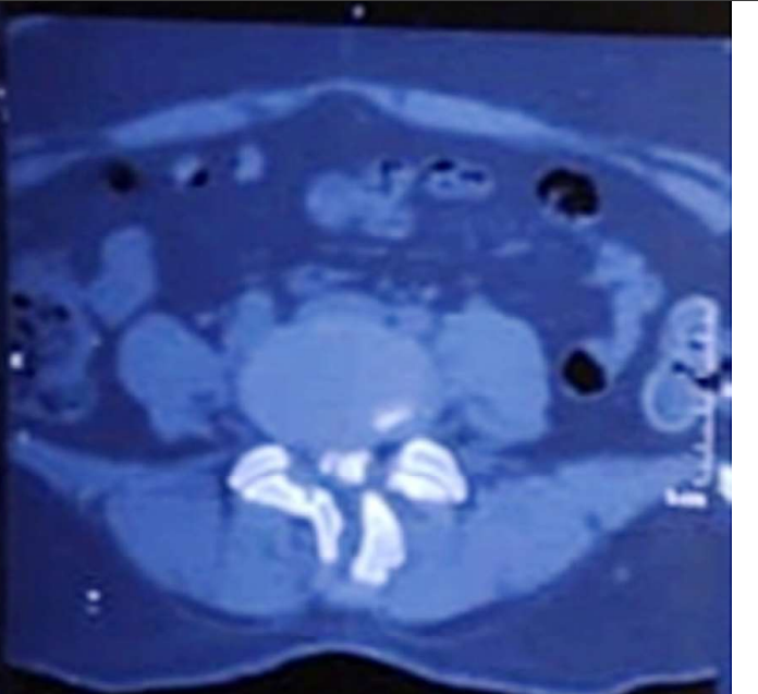
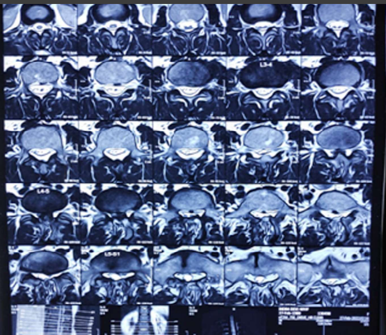
Diastematomyeliais a congenital malformation in which a part of the spinal cord is split, usually at the level of the upper lumbar vertebra in the sagittal direction. It results from an abnormal adhesion between ectoderm and endoderm. Females are affected much more commonly than males (3:1).
There is complete or incomplete sagittal division of the spinal cord into two hemi cords due to the presence of an osseous, cartilaginous or fibrous septum in the central portion of the spinal canal.
The course of the disease is progressive as the patients may be asymptomatic to begin with and gradually progress to sensory motor disorder and loss of bowel and bladder control. With modern imaging techniques, various spinal dysraphism can be diagnosed earlier which can aid in improving quality of life of the individual. Treatment depends upon the symptoms. Regular neurological examinations may helps in early detection of progression of the disease and resection is done if required. We are presenting here a case of diastematomyelia, its imaging study and management.
References
- 1. Doherty D, Walker WO. Neural tube defects. In: Encyclopedia of the Neurological Sciences. 2nd ed. Cambridge, MA: Academic Press; 2014. pp. 360–365.
- 2. Humphreys RP, Hendrick EB, Hoffman HJ. Diastematomyelia. Clin Neurosurg. 1983; 30:436–456.
- 3. Duke Molecular Physiology Institute. Neural tube defects (NTDs). [Accessed Feb 2022]. Shen J, Zhang J, Feng F, Wang Y, Qiu G, Li Z. Corrective surgery for congenital scoliosis associated with split cord malformation: it may be safe to leave diastematomyelia untreated in patients with intact or stable neurological status. J Bone Joint Surg Am. 2016;98:926–936.
- 4. Kaminker R, Fabry J, Midha R, Finkelstein JA. Split cord malformation with diastematomyelia presenting as neurogenic claudication in an adult: a case report. Spine (Phila Pa 1976). 2000; 25:2269–2271.
- 5. Anderson NG, Jordan S, MacFarlane MR, Lovell-Smith M. Diastematomyelia: diagnosis by prenatal sonography. AJR Am J Roentgenol. 1994;163:911–914.
- 6. Arredondo F, Haughton VM, Hemmy DC, Zelaya B, Williams AL. The computed tomographic appearance of the spinal cord in diastematomyelia. Radiology. 1980;136:685–688.
- 7. Han JS, Benson JE, Kaufman B, et al. Demonstration of diastematomyelia and associated abnormalities with MR imaging. AJNR Am J Neuroradiol. 1985;6:215–19.
- 8. Allen LM, Silverman RK. Prenatal ultrasound evaluation of fetal diastematomyelia: two cases of Type I split cord malformation. Ultrasound Obstet Gynecol. 2000;15:78–82.
- 9. Byrd SR, Darling CP, McLone DG. Developmental disorders of paediatric spine. RCNA. 1991;29:4–5.
- 10. Bruberg JA, Latchaw RC, Emanuel K, et al. Magnetic resonance imaging in occult spinal dysraphism. RSNA. 1988;26:181–205.
- 11. Anand AK, Kuchner E, James R. Cervical diastematomyelia: uncommon presentation of a rare congenital disorder. Comput Radiol. 1985;9:45–49.
- 12. Herman TE, Siegel MJ. Cervical and basicranial diastematomyelia. AJR Am J Roentgenol. 1990;154:806–08.
- 13. Castillo M, Mukherji SK. Imaging of paediatric head neck and spine. Philadelphia: Lippincott Raven; 1996. pp. 630–35.
- 14. Banniza von Bazan U, Krastel A, Lohkamp FW. Diastematomyelia - a harmless finding or cause of late neurological disturbance? [Article in German]. Z Orthop Ihre Grenzgeb. 1978;116:72–80.
- 15. Brian EK. Neuroradiology of the spine. In: David S, editor. Textbook of radiology and imaging. 6th ed. New York: Churchill Livingstone; 1998. pp. 1497–99.
Data Sharing Statement
There are no additional data available. All raw data and code are available upon request.
Funding
This research received no funding.
Author Contributions
Whether all authors contributed significantly to the work and approve its publication.
Ethics Declaration
This article does not involve any human or animal subjects, and therefore does not require ethics approval.
Acknowledgements
We would like to express our gratitude to the patients, their families, and all those who have contributed to this study.
Conflicts of Interest
The authors report no conflicts of interest in this work.
About this article
Cite this article
Thanvi S, Singh B. Diastematomyelia: A Case Report. International Journal of Neurology and Neurosurgery. 2023;15(3):99-102.
Licence:
Attribution-Non-commercial 4.0 International (CC BY-NC 4.0)This license enables reusers to distribute, remix, adapt, and build upon the material in any medium or format for noncommercial purposes only, and only so long as attribution is given to the creator.
| Received | Accepted | Published |
|---|---|---|
| July 25, 2023 | September 01, 2023 | October 30, 2023 |
DOI: 10.21088/ijnns.0975.0223.15323.4
Keywords
DiastematomyeliaDysraphismImagingResectionSearch for Similar Articles
Similar Articles
- Intramedullary Capillary Hemangioma of Conus Medullaris: Case Report and Review...
- Atypical Miller Fisher Syndrome
- Therapeutic Potential of Passiflora incarnata in the Management of Neurological...
- Central or Axial Atlantoaxial Dislocation: Redefining Instability at the Craniov...
- Assessment of Neurocognitive Functions in Post-Acute Stroke Patients Undergoing...
Article Level Metrics
Last UpdatedThursday 30 July 2026, 10:11:04 (IST)
1464
Accesses
6
244
00
Citations
NA
NA
NA
Download citation
Article Keywords
Keyword Highlighting
Highlight selected keywords in the article text.
Timeline
| Received | July 25, 2023 |
| Accepted | September 01, 2023 |
| Published | October 30, 2023 |
licence
Attribution-Non-commercial 4.0 International (CC BY-NC 4.0)
This license enables reusers to distribute, remix, adapt, and build upon the material in any medium or format for noncommercial purposes only, and only so long as attribution is given to the creator.