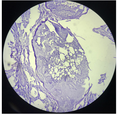
Description: Fig. 1: H & E showing deposition of fat cells in the alveolar parenchyma
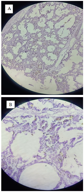
Description: Fig. 2 A & B: H & E micrograph showing deposition of hyaline membrane in the alveoli. A: 10x view, B: 40x view
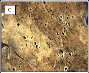
Description: Fig. 2 C: Gross image of hyaline membrane disease
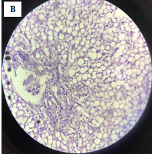
Description: Fig. 3B: H & E micrograph showing extensive mixed (microvesicular and mancrovesicular) fatty changes.
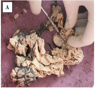
Description: Fig. 3A: Gross image of shrunken liver showing yellowish nodular deposits.
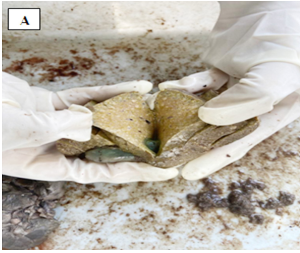
Description: Fig. 4A: Gross image of a single well defined nodule in the brain tissue.
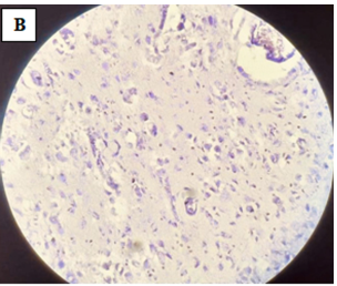
Description: Fig. 4B: H & E micrograph showing large tumor cells (gemistocytes) having abundant eosinophilic cytoplasm and eccentric nuclei
Full-Size
Email
+ Favorites
Export
View in Gallery