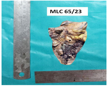
Description: Fig. 1: Heart showing mural clot in the left ventricular cavity
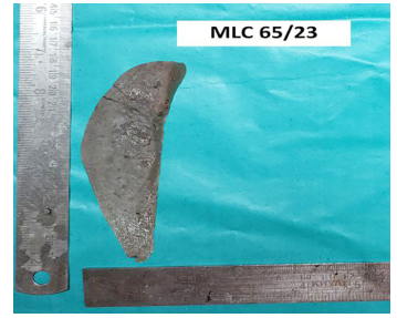
Description: Fig. 2: Splenomegaly
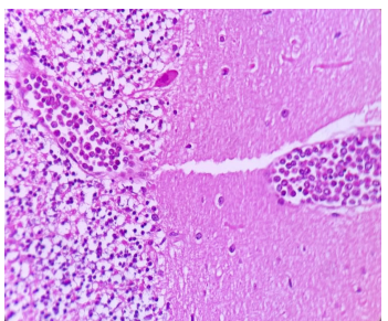
Description: Fig. 3: Cerebellum with neoplastic cells in the blood vessels (H&E,10X)
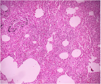
Description: Fig. 4: Lung interstitial infiltrates of neoplastic cells (H&E, 10X)
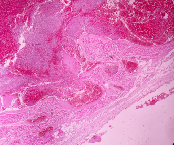
Description: Fig. 5: Mural thrombus with adjacent myocardium (H&E, 40X)
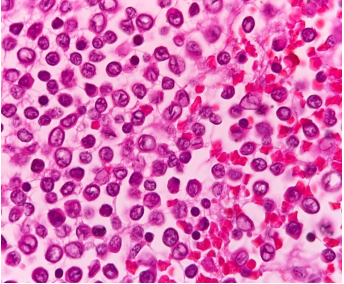
Description: Fig. 6: Blastoid cells exhibiting cleaved nuclei and clumped chromatin (H&E, 100X)
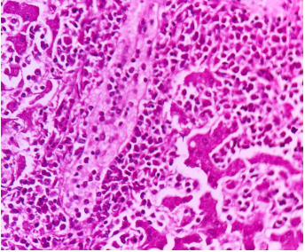
Description: Fig. 7: Liver sinusoids distended and filled with neoplastic cells (H&E,40X)
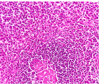
Description: Fig. 8: Spleen with white pulp compressed by surrounding neoplastic infiltrates (H&E, 40X)
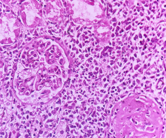
Description: Fig. 9: Kidney showing interstitial neoplastic infiltrate (H&E, 40X)
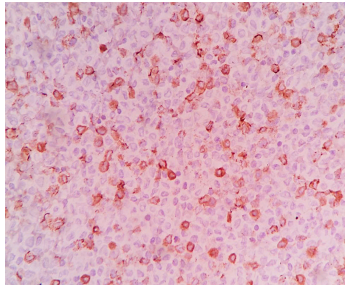
Description: Fig. 10: Neoplastic cells showing diffuse strong granular cytoplasmic positivity for MPO (H&E, 40X)
Full-Size
Email
+ Favorites
Export
View in Gallery