Full Text (PDF)
Indian Journal of Dental Education 16(1):p 39-42, January-March 2023. | DOI: https://doi.org/10.21088/ijde.0974.6099.16123.4
Case Report
Ameloblastic Fibroma Mimicking a Periapical Lesion: A Case Report
Shivani Bansal, Ankita Satish Arvandekar, Neelam N Andrade, Rajiv S. Desai, Pooja Prasad
Author Information
Licence:
Attribution-Non-commercial 4.0 International (CC BY-NC 4.0)This license enables reusers to distribute, remix, adapt, and build upon the material in any medium or format for noncommercial purposes only, and only so long as attribution is given to the creator.
Indian Journal of Dental Education 16(1):p 39-42, January-March 2023. | DOI: https://doi.org/10.21088/ijde.0974.6099.16123.4
How Cite This Article:
Arvandekar AS, Bansal S, Andrade NN, et al. Ameloblastic Fibroma Mimicking a Periapical Lesion: A Case Report. Indian J Dent Educ. 2023;16(1):39-42.Timeline
Received : October 15, 2022
Accepted : November 08, 2022
Published : November 30, 2022
Abstract
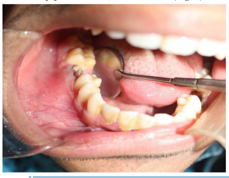
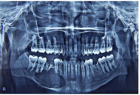
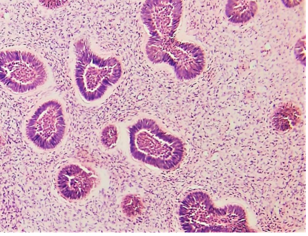
Ameloblastic fibroma (AF) is an uncommon true mixed odontogenic tumor, frequently diagnosed in the first two decades of life and presented as a well defined unilocular or multilocular radiolucency. We report an unusual case of AF mimicking a periapical lesion in a 22‑year‑old male as a painless swelling associated with a carious molar. The patient underwent surgery and the lesion was excised, subsequently submitted for histopathological examination. Thus, this paper highlights the importance of submission of periapical tissue for its histopathological examination.
References
- 1. Muller S, Vered M, Takata T, Slootweg PJ. Odontogenic and maxillofacial bone tumours. In: El-Naggar AK, Chan JKC, Grandis JR, Takata T, Slootweg PJ, editors. WHO classification of Head and Neck tumours. 4th ed. Lyon: IARC; 2017. p. 222-223.
- 2. Dahlkemper P, Wolcott JF, Pringle GA, Hicks ML. Periapical central giant cell granuloma: A potential endodontic misdiagnosis. Oral Surg Oral Med Oral Pathol Oral Radiol Endod. 2000;90(6):739-745.
- 3. Nary FH, Matsumoto MA, Fraga SC, et al. Periapical radiolucency mimicking an odontogenic cyst. Int Endod J. 2004;37(5):337–344.
- 4. Reichart PA, Philipsen HP. Ameloblastic fibroma. In: Reichart PA, Philipsen HP, editors. Odontogenic Tumours and Allied Lesions. London: Quintessence; 2004. p. 121-129.
- 5. Gupta S, Tandon A, Mehrotra D, Gupta OP. Ameloblastic fibroma: Report of 3 cases and literature review. Int J Oral Maxillofac Pathol. 2011;2:59-63.
- 6. Tozoglu S, Hatipoglu M, Aytekin Z, Gurer EI. Extensive ameloblastic fibroma of the mandibula in a female adult patient: A case report with a follow-up of 3 years. Eur J Dent. 2016;10(1):139-143.
- 7. Soluk-Tekkesin M, Wright JM. The World Health Organization Classification of Odontogenic Lesions: A Summary of the Changes of the 2017 (4th) Edition. Turk Patoloji Derg. 2018;34(1):1-18.
- 8. Kumar RM, Bavle R, Srinath Z, Umashankar DN. Ameloblastic fibroma in a young adult. J Oral Maxillofac Pathol. 2019;23(Suppl 1):S63-S65.
- 9. Zallen RD, Preskar MH, McClary SA. Ameloblastic fibroma. J Oral Maxillofac Surg. 1982;40(8):513-517.
- 10. Trodahl JN. Ameloblastic fibroma. A survey of cases from the Armed Forces Institute of Pathology. Oral Surg Oral Med Oral Pathol. 1972;33(4):547-558.
- 11. Slootweg PJ. An analysis of the interrelationship of the mixed odontogenic tumors: ameloblastic fibroma, ameloblastic fibro-odontoma, and the odontomas. Oral Surg Oral Med Oral Pathol. 1981;51(3):266-275.
- 12. Kashyap B, Reddy PS, Desai RS. Plexiform ameloblastoma mimicking
Data Sharing Statement
There are no additional data available. All raw data and code are available upon request.
Funding
This research received no funding.
Author Contributions
Whether all authors contributed significantly to the work and approve its publication.
Ethics Declaration
This article does not involve any human or animal subjects, and therefore does not require ethics approval.
Acknowledgements
We would like to express our gratitude to the patients, their families, and all those who have contributed to this study.
Conflicts of Interest
The authors report no conflicts of interest in this work.
About this article
Cite this article
Arvandekar AS, Bansal S, Andrade NN, et al. Ameloblastic Fibroma Mimicking a Periapical Lesion: A Case Report. Indian J Dent Educ. 2023;16(1):39-42.
Licence:
Attribution-Non-commercial 4.0 International (CC BY-NC 4.0)This license enables reusers to distribute, remix, adapt, and build upon the material in any medium or format for noncommercial purposes only, and only so long as attribution is given to the creator.
| Received | Accepted | Published |
|---|---|---|
| October 15, 2022 | November 08, 2022 | November 30, 2022 |
DOI: https://doi.org/10.21088/ijde.0974.6099.16123.4
Keywords
Ameloblastic fibromaOdontogenic cystsOdontogenic tumorsPeriapical lesionSearch for Similar Articles
Similar Articles
- Post Extraction Surgical Ciliated Cyst of Maxilla: Case Report of Unusual Pathog...
- Natural Tooth Surgical Stent (NTSS) Technique For Prosthodontically Driven Rehab...
- Growth Meets Diagnosis: Revisiting Synchondrosis in the Orthodontic Era
- The Silent Struggle to Breathe: A Review on Obstructive Sleep Apnea
- Evaluating Awareness of Oral-Systemic Health Interactions in Urbanized Indian So...
Article Level Metrics
Last UpdatedWednesday 17 June 2026, 23:10:36 (IST)
1363
Accesses
8
435
00
Citations
NA
NA
NA
Download citation
Article Keywords
Keyword Highlighting
Highlight selected keywords in the article text.
Timeline
| Received | October 15, 2022 |
| Accepted | November 08, 2022 |
| Published | November 30, 2022 |
licence
Attribution-Non-commercial 4.0 International (CC BY-NC 4.0)
This license enables reusers to distribute, remix, adapt, and build upon the material in any medium or format for noncommercial purposes only, and only so long as attribution is given to the creator.