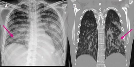
Description: Fig. 1: (A) Chest X-Ray showed multiple radio opacities in bilateral lung fields, (B) CT chest showed Patchy subpleural consolidations with nodules in bilateral lungs
Heading
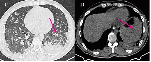
Description: Fig. 2: (C) CT chest showed Patchyperibronchialconsolidations with nodules in bilateral lungs, (D) CT abdomen showed few hypodense lesions spleen suggestive of micro-abscesses
Heading
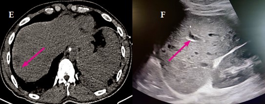
Description: Fig. 3: (E) CT abdomen showed few hypodense lesions in liver suggestive of micro-abscesses, (F) Ultrasound abdomen showed few micro-abscesses in spleen
Heading
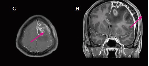
Description: Fig. 9.4: (G) MRI brain with contrast showed Conglomerate peripheral rim enhancing lesions in left frontal lobe, (H) Multiloculated extra-axial collection with dural enhancement
Heading
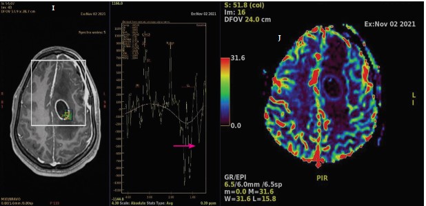
Description: Fig. 5: (I) MR spectroscopy revealed large lipid-lactate peaks within the lesion (J) MR perfusion showed no increase in perfusion