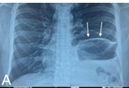
Description: (A) Plain X-ray chest showing herniated contents thru left BH(white arrows),
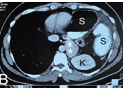
Description: (B) CECT chest axial view herniated stomach (‘S’), left kidney (‘K’), spleen (white asterisks) & pancreas(black asterisk),
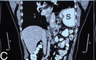
Description: C) CECT oblique view herniated stomach, spleen(white asterisk) & pancreas (black asterisks)
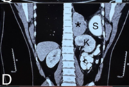
Description: (D) CECT coronal view showing herniated stomach, kidney and spleen(black asterisk),
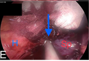
Description: (E) Operative pic showing lysis of small bowel adhesions to parietes (blue arrow)
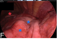
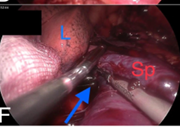
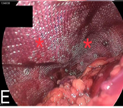
Description: (F) shows view of left chest thru BH with large stretch of stomach (blue asterisks) & left lung (red asterisk)
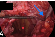
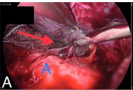
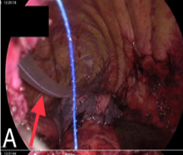
Description: (A) shows left chest cavity thru BH(red asterisk) & lysis of lateral chest wall adhesions
Description: (B) Lysis of adhesions betn stomach & diaphragm
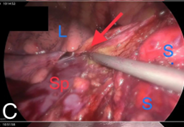
Description: (C) Lysis of adhesions betn spleen & stomach(red arrow) which are in volvulus, also seen is lower part of left lung
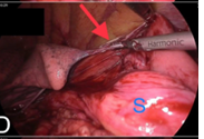
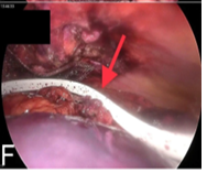
Description: (D) Lysis of adhesions betn lung & stomach(red arrow),
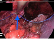
Description: (E) Lysis of adhesions betn heart & spleen(blue arrow)
Description: (F) Lysis of adhesions betn upper pole of spleen & surrounding structures(blue arrow)
Description: (A) Lysis of adhesions betn spleen & thoracic aorta,
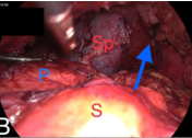
Description: (B) Mobilised spleen, pancreas & stomach after adhesiolysis & correction of volvulus,
Description: (C) Delivery of spleen (red asterisks) & stomach (blue asterisks) into the abdomen frm left chest
Description: (D) Lysis of adhesions betn left kidney(blue asterisks) & surroundings (red arrow)
Description: (E) Assessment of large muscle defect for closure (blue arrow)
Description: No description available.
Description: A) Left ICD inserted (red arrow)
Description: B, C) Suturedarning performed after inability to close defect,
Description: D & E) Mesh (red asterisks) placed over the defect, thereafter & tack-fixed to the edges of the defect
Description: (F) Flat drain placed in situ (red arrow)
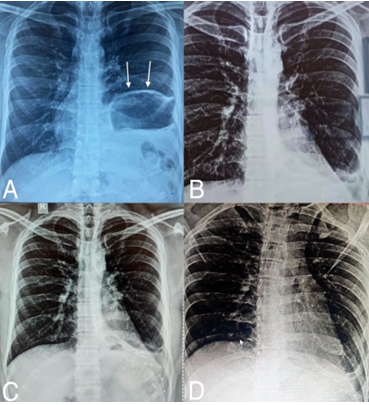
Description: Serial X-ray Chest films. A) Preopfilm showing Lt BH (white arrows), B, C, D) 1, 3 & 5 months postop films respectively, showing left costo-phrenic angle getting progressively clearer
Full-Size
Email
+ Favorites
Export
View in Gallery