Description: Shows central cyanosis evident in the lips of an uncorrected TOF
Heading
Description: Illustrates Pandigital clubbing in an uncorrected TOF
Heading
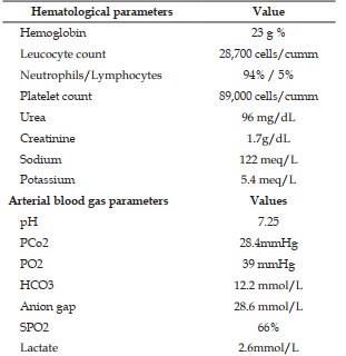
Description: Various blood investigations obtained on arrival to ED
Heading
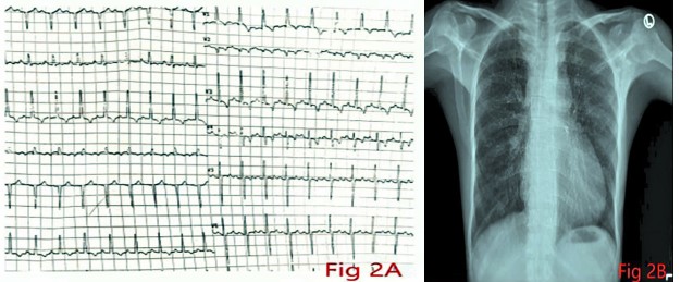
Description: Figure 2A: Electrocardiogram showing sinus tachycardia of HR 144 bpm, right axis deviation, and RBBB. Also note the positive R wave in V1, suggesting pulmonary hypertension. Figure 2B: Chest X-ray showing pulmonary oligemia in an uncorrected TOF
Heading
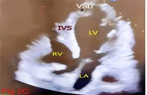
Description: Shows an echocardiography image of the index patient revealing a large VSD
Heading
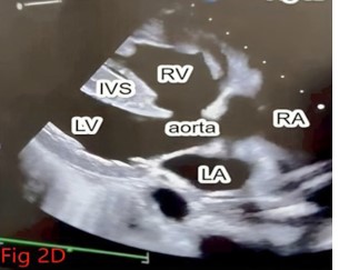
Description: Shows an echocardiography image of the index patient showing an overriding aorta arising from both ventricles. RA - Right atrium, LA - Left atrium, RV - Right Ventricle, LV - Left ventricle, Ivsinterventricular septum