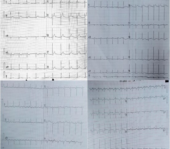
Showing ECG of the respective case, Showing flattening of the T wave( arrows).
Description: No description available.
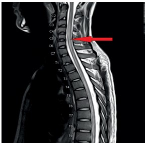
case 1-mri (non-contrast) cervicodorsal spine showing sagittal and axial planes of T2 weighted images indicating hyperintense lesion extending from c3 level to d10 level (indicated by red arrow)
Description: No description available.
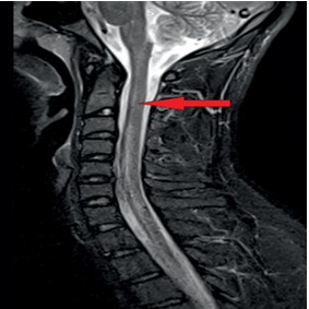
case 3-Long segment intra-medullary hyperi ntensity occupying more than 2/3rd of the cord diameter is noted from C6-7 level to D4-5 level – suggestive of longitudinally extensive transverse myelitis (indicated by red arrow).