Full Text (PDF)
RFP Journal of ENT and Allied Sciences 9(2):p 97-101, July-December 2024. | DOI: n.a
Case Report
A Spontaneous Middle Ear Encephalocele with Unilateral Chronic Otitis Media
Author Information
Licence:
Attribution-Non-commercial 4.0 International (CC BY-NC 4.0)This license enables reusers to distribute, remix, adapt, and build upon the material in any medium or format for noncommercial purposes only, and only so long as attribution is given to the creator
RFP Journal of ENT and Allied Sciences 9(2):p 97-101, July-December 2024. | DOI: n.a
How Cite This Article:
Gupta K. A spontaneous middle ear encephalocele with unilateral chronic otitis media. RFP J ENT Allied Sci. 2024;9(2):97–102.Timeline
Received : November 23, 2024
Accepted : December 15, 2023
Published : December 30, 2024
Abstract
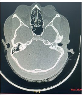
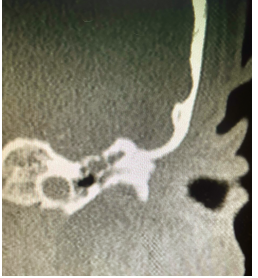
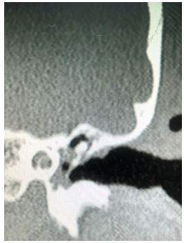
Introduction: Heterotopic Brain and Encephaloceles of middle ear usually present with symptoms of ear discharge and hearing loss. Some patients have additional symptoms of headache, abnormal ringing sensation and vertigo. Radiology may or may not show a communication with central nervous system in encephaloceles and the bony defect may be unnoticeable or attributed to the thinning of bone due to chronic otitis media. On computerised tomography, no distinction may be made between fluid, granulations, cholesteatoma and other space occupying lesions. Morphologically, it may be identified grossly if it presents with its characteristic pink colour and typical convoluted cerebriform pattern (CCP). Methods: A 26 years female presented to us with unilateral ear discharge since birth and unilateral hearing loss on ipsilateral side for last 18 months. Clinically, radiologically and morphologically it mimicked chronic otitis media with cholesteatoma with no pre-operative and intra-operative distinction from the later. However, the coronal section at the level of anterior tegmen showed a small defect of the tegmen. T2 weighted MRI confirmed presence of brain tissue in middle ear and mastoid cavity. Results: Combined Middle Cranial Fossa and mastoid approach were employed, unviable brain tissue was excised and sent for histopathology. Viable brain tissue was reduced and tegmen defect was closed with temporalis muscle, conchal cartilage and temporalis fascia. Histopathology revealed glial tissue, ependyma and choroid plexus without any cholesteatoma. Conclusion: Tympanic encephalocelesare very rare and may closely mimic chronic otitis media. It may become very difficult to differentiate between the two which may lead to unexpected complications andloss
References
- 1. Farneti P., Balbi M. and Foschini M.P. Neuroglial Choristoma of the Middle Ear. Acta Otorhinolaryngol Ital 2007; 27(2):94-7.
- 2. Gyure K.A., Thompson L.D., Morrison AL. A clinicopathological study of 15 patients with neuroglial heterotopias and encephaloceles of the middle ear and mastoid region. Laryngoscope 2000;110:1731-5.
- 3. Devaney KO, Rinaldo A, Ferlito A. Teratoma of the Middle Ear: A real entity or a non-entity? Acta Otolaryngol 2005; 125:122-4.
- 4. El Hadi, Sorrentino T, Calmels MN, Fraysse B, Deguine O, Marx M. Spontaneous Tegmen Defect and Semicircular Canal Dehiscence: Same Etiopathogenic Entity? OtolNeurotol 2012; 33:591-5.
- 5. Baron SH. Herniation of the Brain into the Mastoid Cavity Postsurgical, Postinfectional, or Congenital. Arch Otolaryngol 1969;90(6):779-85.
- 6. Schurr PH. Endaural Cerebral Hernia. Br J Surg1960; 47:414-7.
- 7. Blatt IM. Surgical Repair of Cerebrospinal Otorrhea due to Middle Ear and Mastoid DiseaseLaryngoscope 1963; 73:446-60.
- 8. Mealey J Jr. Chronic Cerebrospinal Fluid OtorrheaNeurology 1961; 11:996-8.
- 9. Plontke SK, Preyer S, Pressler H, Mundinger PM, Plinkert PK. Glial lesion of the infratemporal fossa presenting as a soft tissue middle ear mass – rudimentary encephalocele or neural crest remnant? Int J Pediatr Otorhinolaryngol 2000; 56:141-7.
- 10. Vasama JP, Ramsay H, Markkola A. Choristoma of the middle ear. OtolNeurotol 2001; 22:421-2.
- 11. Heffner DK. Brain in the middle ear or nasal cavity: heterotopia or encephalocele? Ann DiagnPathol 2004; 8:252-7.
- 12. Juliano AF. Cross Sectional Imaging of the Ear and Temporal Bone. Head Neck Pathol 2018:12(3):302-20.
- 13. Vvan de Graaf FW, Lange MM, Spakman JI, van Grevenstein WMU, Lips D, de Graaf EJR et al., (2019) Comparison of systematic video documentation with narrative operative report in colorectal cancer surgery. JAMA Surgery 154(5):381–389.
- 14. Van de Graaf FW, Lange MM, Menon AG, O’Mahoney PR, Milsom JW, Lange JF (2016) Imaging for quality control: comparison of systematic video recording to the operative note in colorectal cancer surgery. A pilot study. Ann Surg Oncol 23:798–803
Data Sharing Statement
There are no additional data available
Funding
This research received no funding
Author Contributions
All authors contributed significantly to the work and approve its publication
Ethics Declaration
This article does not involve any human or animal subjects, and therefore does not require ethics approval
Acknowledgements
Information Not Provided
Conflicts of Interest
No conflicts of interest in this work
About this article
Cite this article
Gupta K. A spontaneous middle ear encephalocele with unilateral chronic otitis media. RFP J ENT Allied Sci. 2024;9(2):97–102.
Licence:
Attribution-Non-commercial 4.0 International (CC BY-NC 4.0)This license enables reusers to distribute, remix, adapt, and build upon the material in any medium or format for noncommercial purposes only, and only so long as attribution is given to the creator
| Received | Accepted | Published |
|---|---|---|
| November 23, 2024 | December 15, 2023 | December 30, 2024 |
DOI: n.a
Keywords
Encephalocele Congenital Middle Ear Heterotopic Brain Cholesteatoma Cranial Fossa Combined Approach Surgery.Search for Similar Articles
Similar Articles
- Pseudomonas Aeruginosa in an Immunocompetent Individual with Acute Tonsillitis;...
- Hidden Dangers of Oral Hygiene: Oropharyngeal Impalement by Toothbrush in a 3-Ye...
- Functional Outcome of Abbe–Estlander Flap in Lower Lip Carcinoma with Commissura...
- Primary Laryngeal Histoplasmosis
- Inverted Papilloma a Retrospective Study of 17 Cases
Article Level Metrics
Last UpdatedMonday 26 January 2026, 20:20:38 (IST)
149
Accesses
1
30
00
Citations
NA
NA
NA
Download citation
Article Keywords
Keyword Highlighting
Highlight selected keywords in the article text.
Timeline
| Received | November 23, 2024 |
| Accepted | December 15, 2023 |
| Published | December 30, 2024 |
licence
Attribution-Non-commercial 4.0 International (CC BY-NC 4.0)
This license enables reusers to distribute, remix, adapt, and build upon the material in any medium or format for noncommercial purposes only, and only so long as attribution is given to the creator