Heading
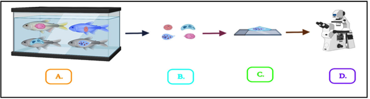
Description: Fig. 1: In this schematic diagram, fishes were infected with different parasites in an aquaculture tank (A), isolation the parasites were gently dissected (B), the fresh smear was made grease free slide with the help of water (C) and finally observed under microscope at different magnifications (D).