Heading
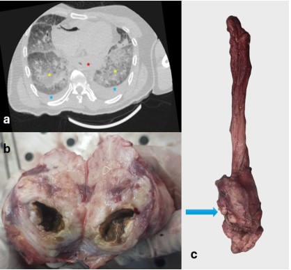
Description: A: Postmortem CT axial section in lung window showing a hyperdense mass at the lower end of the
esophagus (Red Asterix), bilateral pleural effusion (Blue Asterix) and bilateral ground-glass opacities along with
interlobular septal thickening (Yellow Asterix); B: The cut section of esophageal mass showing irregular and uneven
margins, and areas of necrosis; C: showing a mass located at the lower end of the esophagus (Blue arrow).