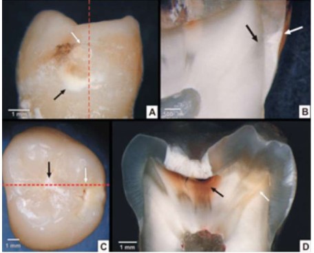
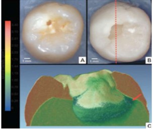
Description: (A&B) First detectable change in dentin in response to cariogenic biofilm is an area of sclerotic dentin subjacent to demineralized enamel, (C&D) Yellow to dark-brown discolouration of progressed carious lesion along DEJ
Heading
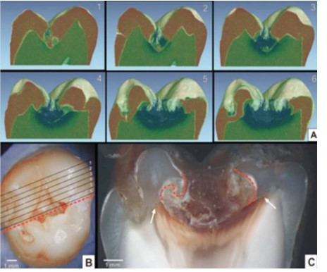
Description: (A&B) Different phases of caries progression within one lesion, (B&D) “hidden” caries lesion
Heading
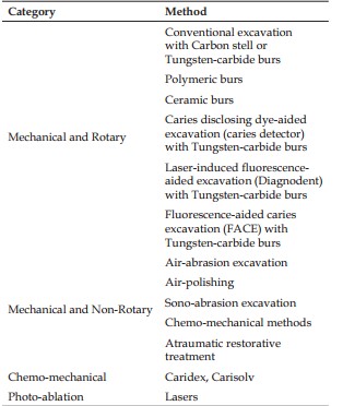
Description: Classification of Caries Excavation Methods (According to Banerjee et al. 2000):12
Heading
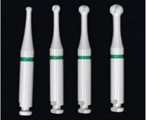
Description: CeraBurs with different diameters. From left to right: 10, 14, 18, and 23-mm diameter
Heading
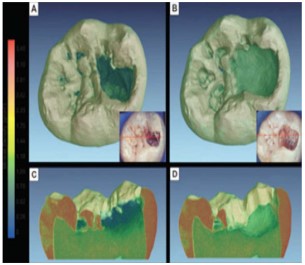
Description: Caries excavated using ceramic burs in 3D volumetric reconstruction of carious tooth
Heading
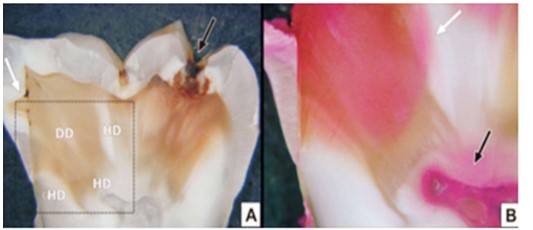
Description: A: Mesiodistal section of a tooth presenting occlusal dentin caries (black arrow) and an approximal lesion (white arrow). DD = demineralized dentin; HD=hypermineralized dentin. B: Higher magnification of the dotted area in Fig A after staining with a 1% acid red-in-propylene-glycol solution. Note staining of circumpulpal dentin (black arrow) and areas of
hypermineralized dentin (white arrow).
Heading
Description: Volume-rendering slices based on micro-CT evaluation data of carious tooth before and after caries excavation aided
by prototype plastic instrument with SFC-VIII
Heading
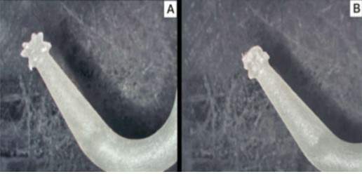
Description: Prototype plastic instrument (Star v 1.8, 3M ESPE) (A) before and (B) after caries
Heading
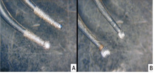
Description: (A) Diamond-coated tips with different diameters for enamel preparation, (B) Tungstencarbide tips with different diameters for dentin excavation