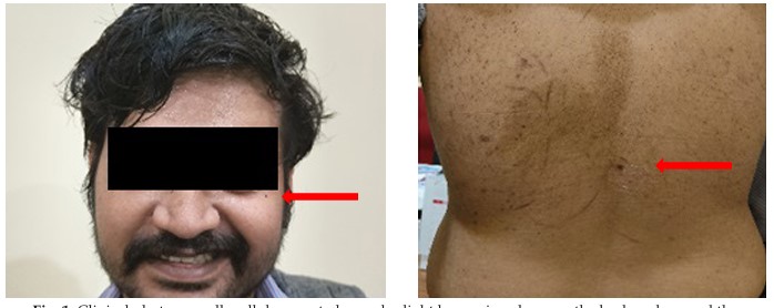
Clinical photo – small well demarcated macules light brown in color over the back and around the eyes (red arrow)
Description: No description available.
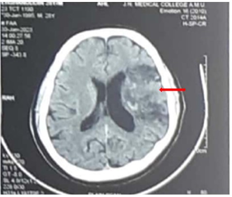
CT Brain – Image suggestive of sub-acute infarct (red arrow) involving left fronto-parietal-temporal lobe with gyriform hyperdensities with CT value of blood noted within the infarct (hemorrhagic transformation)
Description: No description available.
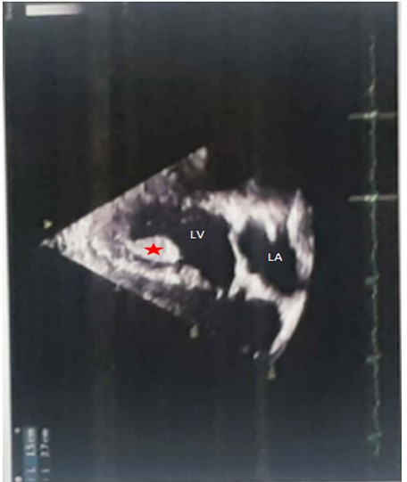
ECHO – A mass of size 2.7 x 1.5 cm is seen attached to the LV apex with a stalk with a high embolic potential (red asterix). Regional wall motion abnormality present in the LAD territory. No chamber enlargement and all valves are within normal limits. LV – Left ventricle, LA – Left atrium
Description: No description available.
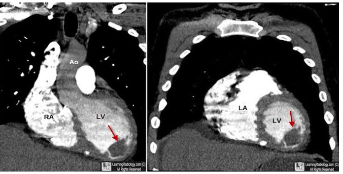
MRI cardiac –A 3.2 x 2.5 cm pedunculated mass seen arising from the LV apex with with heterogenous signal enhancement in T1 and T2 sequences (red arrow) (Source: https://learningradiology.com/notes/ cardiacnotes/leftventthrombus.htm)
Description: No description available.
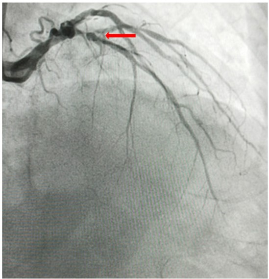
CAG - A coronary angiogram showing an ostio-proximal lesion of LAD/D1 around 80-90% with diffusely diseased LAD (red arrow)
Description: No description available.
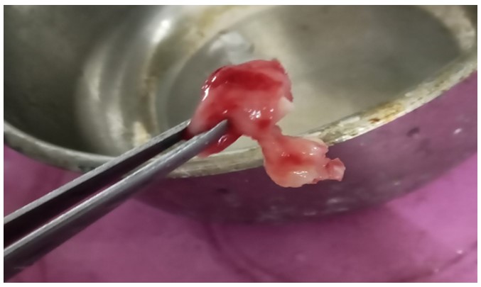
Surgical specimen – A 3 x 2 cm fleshy reddish mass excised from the apex of the left ventricle.
Description: No description available.
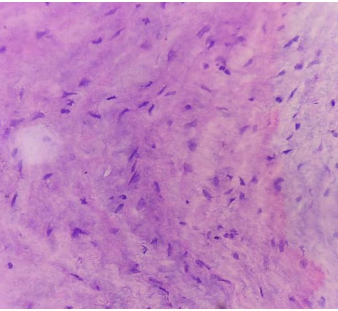
HPE - H and E stained section shows stellate to spindle cells with eosinophilic cytoplasm with indistinct border within myxoid stroma.