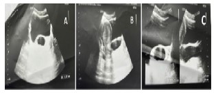
A-C: Ultrasound TAS image showing a well defined longitudinal cystic lesion of 7.34(CC)×2.24(AP)×3.37(TR) in anterior vaginal wall with most probability of being gartner duct cyst
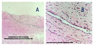
3 A-B: A - showing cyst with fibrocollagenous tissue and muscle bundles on H&E at 40X magnification, B - cuboidal epithelium lining the cyst wall on H&E at 400X magnification