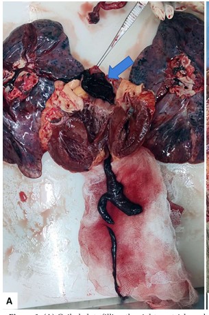
) Coiled clots filling the right ventricle and extending into the pulmonary arteries of both lungs (saddle embolus), along with clots retrieved from the inferior vena cava and common iliac vein
Description: No description available.
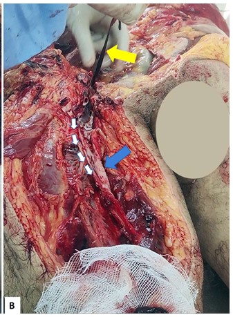
Dissected right femoral vein (blue arrow) with adherent clots being lifted (yellow arrow) from the vessel during autopsy.
Description: No description available.
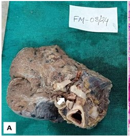
Formalin-fixed lung specimen showing a clot within the pulmonary artery (white arrow indicates a clot extending from the right ventricle
Description: No description available.
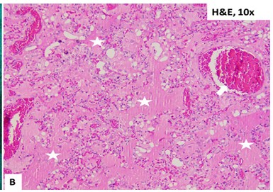
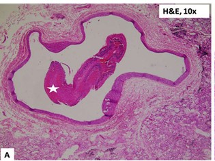
Histologic section of lung parenchyma (H&E stain) showing extensive pulmonary edema (white star) and congested blood vessels (white arrow), with areas of interstitial hemorrhage.
Description: No description available.
Histologic section of a hilar pulmonary vessel showing an occlusive blood clot (white star)
Description: No description available.
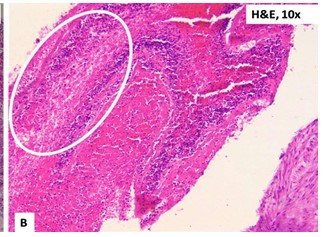
Cross section of a thrombus showing distinct dark-red layers (rich in red blood cells) and pale layers (fibrin and platelets), demonstrating the lines of Zahn indicative of an antemortem thrombus (white oval highlight)