Pathophysiological cascade involved in Wolf's isotopic response
Description: No description available.
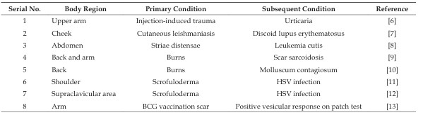
Clinical image depicting erythematous plaque (3 cm x 2cm) with minimal scaling, present over dorsum of right hand, over an atrophic burn scar
Description: No description available.
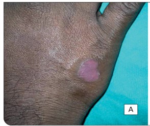
Dermoscopy (DinoLite II, Pro HR dermatoscope) at 10x magnification, in polarized mode revealed, regularly arranged red dots indicative of dotted vessels, on diffuse pink background
Description: No description available.
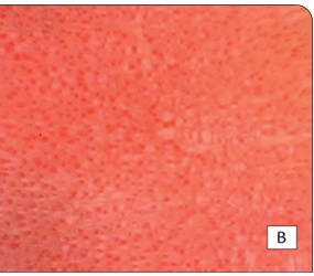
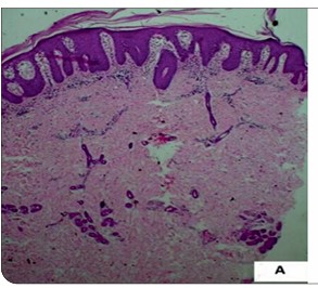
Regular acanthosis with club-shaped rete ridges and superficial dermal, perivascular infiltrate (H & E, 4X),
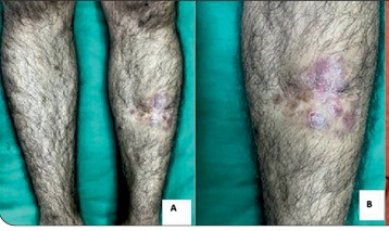
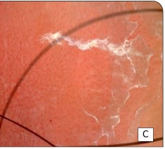
Clinical image depicting multiple erythematous plaques ranging in size from 1.5 cm x 1 cm to 3 cm x 2cm with minimal scaling, present over left lower shin, overlying an atrophic post- traumatic scar, Polarised dermoscopy (DinoLite II pro HR dermatoscope) at 10x magnification highlighted regularly arranged red dots,
Description: No description available.
Reminiscent of dotted vessels, on diffuse pink background
Description: No description available.
] Regular acanthosis with club-shaped rete ridges and superficial dermal, perivascular infiltrate (H & E, 4X)
Description: No description available.
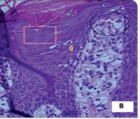
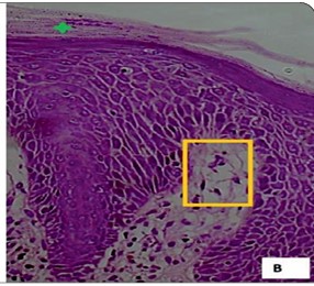
Munro’s microabscess (green star) and dilated papillary dermal blood vessels with perivascular infiltrate (yellow rectangle) (H &E, 40 X).